Biomedical Engineering Reference
In-Depth Information
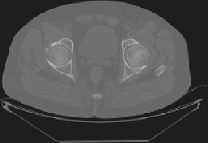
Fig. 2
The transverse section of total femur CT image
scanning of patients are obtained using GE Ultrafast High-resolution Multislice CT
Scanner (16 Slice) containing a total number of 909, 667, and 1,714 images,
respectively, pixel size of 0.7031, 0.8867, and 0.9766 mm, respectively, slice
thickness of 0.4 mm, and resolution of 512 9 512. DICOM file is a standard for
handling, storing, printing, and transmitting information in medical imaging and
contains binary data elements. In MIMICS, distinctive CT images are a pixel map of
the linear X-ray attenuation coefficient of tissue. The pixel values are scaled so that
the linear X-ray attenuation coefficient of air equals -1024 and that of water equals 0.
This scale is called the Hounsfield scale after Godfrey Hounsfield, one of the pioneers
in computerized tomography. Using this scale, fat is around -110, muscle is around
40, trabecular bone is in the range of 100-300, and cortical bone extends above
trabecular bone to about 2,000. The pixel values are shown graphically by a set of
gray levels that vary linearly from black to white [
20
].
Image Segmentation
Three models of right proximal human femur of three individual male human
patients are created: Model 1 of 17-year-old male, Model 2 of 32-year-old male,
and Model 3 of 40-year-old male. MIMICS is an interactive tool for the visuali-
zation and segmentation of CT images as well as MRI images and 3D rendering of
objects. Therefore, in the medical field MIMICS is used for diagnostic, operation
planning, or rehearsal purposes. Figure
2
shows the image of normal individual
total femur as acquired from the DICOM file images after conversion in MIMICS
v10. Bone tissue is then extracted by means of thresholding using default values
range. The extracted bone tissue is put into a mask. Mask is a collection of pixels
which can be modified using various tools successively: edit masks, region
growing, and calculate of 3D mask. Edit mask is used to separate the whole total
femur from the adjoining hard tissues like pelvis, tibia, etc. The region growing
Search WWH ::

Custom Search