Biomedical Engineering Reference
In-Depth Information
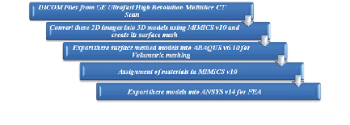
Fig. 1
Steps involved in finite element modeling
replacement of joint in Indian subjects, as these properties are needed before finite
element analysis of indigenized hip joint to study its stability in the bone [
17
,
18
].
The role of ante version in transferring the load from implant to bone and its influence
on total hip arthroplasty (THA) is determined. Also, loading of the proximal femur
during daily activity, i.e., walking and stair climbing is determined. Experimental
and analytical approaches are used to determine the in vivo loading of the hip joint.
A numerical muscular skeleton model is validated against measured in vivo hip
contact force [
13
,
19
].
Materials and Methods
A biomechanical analysis of human femur under half the body weight which varies
with the shift of the vertical axis of body center where center of gravity of the body
lies as one leg is lifted in air while the other leg is on the ground. This condition of
instability is essential for the understanding of failure mechanisms during day-to-day
life conditions or any accidental cases, as with every half-centimeter shift of this
vertical axis of body center 5 kg weight is added to the body weight acting on the leg
touching the ground, for providing guidance for the design and operation of human
femur replacement. 3D FE models are created using CT images in materialized
interactive medical image control system (MIMICS) and the various steps involved
are described in Fig.
1
. For biomechanical investigation of total deformation, stress
distribution, and fatigue tools throughout the right proximal human femur of all male
patients, under physiological loading conditions, FEA is performed.
Image Acquisition
The CT data of total femur of normal individual male patients of 17, 32, and 40 years
are collected. This geometrical data of real proximal human femur bone are in the
form of digital imaging and communications in medicine (DICOM) files. The CT
Search WWH ::

Custom Search