Biomedical Engineering Reference
In-Depth Information
c) Cast from MSPs 3
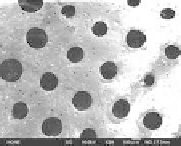
a) Cast from MSPs 1
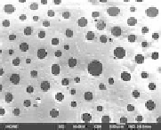
b) Cast from MSPs 2
Figure 8.19
Surface coniguration of PLCL scaffolds cast from MSPs 1 (295
± 105
mm), MSPs 2 (369
±
59
μ
m) and MSPs 3 (456 ± 24
μ
m).
Small pores less than 20
μ
m were generated from co-existing
phase of PLCL, chloroform, and hexane.
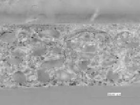
Interior coniguration of scaffolds was observed to conirm 3D
porous structure by a SEM and a stereomicroscope (Fig. 8.20). Figure
20(b) synthesized from 2D images captured at seven different focal
depths using VHX-900. Therefore, the coniguration of the scaffold
to the direction of depth can be observed very well. Well-connected
pores owing to magnet-assisted assembly of MSPs and successful
iniltration of polymer can be conirmed from these images.
a)
b)
500
m
Figure 8.20
Interior coniguration of sheet-like scaffolds captured by (a)
SEM, and (b) stereomicroscope.
8.7.3
Evaluation of Young's Modulus and Porosity
Strength and compliance of sheet-like scaffolds were evaluated in
terms of Young's modulus and porosity. Those parameters are very
important for scaffolds to maintain their shape and it to surrounding
tissue.
8.7.3.1
Evaluation of Young's modulus
Tensile tests were performed to measure Young's modulus of sheet-
like scaffolds fabricated in the previous section and nonporous PLCL.


Search WWH ::

Custom Search