Biomedical Engineering Reference
In-Depth Information
P
5
was successfully detected by the system to keep tracking the
catheter. The most probable reason is the change in the vascular
lumen shape, because
P
5
was placed away from the acrylic frame
holding the silicone model. For this application, the detection range
could be increased up to 15 mm to give more time to the robotic
camera to set. A larger detection range between
P
3
and
P
4
will
produce ambiguities in
M
0
.
7.3.5
Integration of the Robotic Camera with EVE
Integrating EVE to the robotic camera enabled to perceive the
silicone vasculature on the same way that human vasculature is
perceived during an endovascular intervention. Improving the
quality of the training system as the user will see the catheter
through a video monitor as surgeons does in endovascular surgery.
An optional control with joystick enabled to operate the camera
as the luoroscope is manipulated in IVR. But depending on the
training purpose an automatic motion of the camera and DSA image
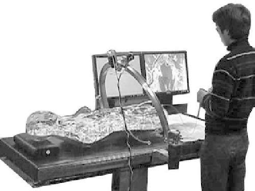
generation is desired. As shown in Fig. 7.10, the vasculature is
visible in all moments during the simulation procedure. To make the
process more realistic is necessary that silicone vasculature is visible
only when a contrast media is injected or after the application DSA
process.
Robotic Camera
EVE
Figure 7.10
Endovascular intervention simulation with EVE and robotic
camera.


Search WWH ::

Custom Search