Biomedical Engineering Reference
In-Depth Information
EXAMPLE PROBLEM 6.5
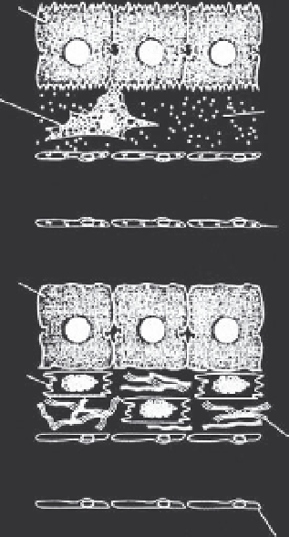
The hepatic stellate cell is a mesenchymal cell that is located in the Space of Disse, the area
between the sheets of hepatocytes and the hepatic sinusoidal endothelium (Figure 6.26) in the liver.
In the normal state, hepatic stellate cells are found in close proximity to a basement membrane-like
matrix. This matrix consists of collagen type IV, laminin, and heparan sulphate proteoglycans. If the
liver is injured, hepatic stellate cells are activated, and they begin to make matrix proteins, mostly
collagen type I and III. As a result of hepatic stellate cell activation, the phenotype of both the hepa-
tocytes (lose their brush border) and endothelium (lose their fenestration) changes. If this condition
persists, liver fibrosis results. Thus, a lack of coordination in cellular function in the tissue micro-
environment can lead to loss of tissue function. This example can be used to examine how liver
dysfunction can be prevented or corrected, including using a tissue engineering approach.
Hepatocyte
Quiescent
HSC
Basement
membrane-like
matrix
Sinusoidal lumen
Fenestrated
endothelium
Hepatocyte
Activated
proliferative
HSC
Matrix rich in
Interstitial
collagens I
and III
Sinusoidal lumen
Endothelial
cells lose
fenestrae
FIGURE 6.26
Schematic of hepatic stellate cell activation and the process of liver fibrosis.
From Iredale, 1997.
Tissues are perfused by the microcirculation and can be viewed as essentially homoge-
neous down to a length scale of about 100
m. The microcirculation then interfaces with
the long-distance convective transport system that connects all the tissues in the body
and, thus, all cells, to a nutritional supply, exchange of respiratory gases, removal of toxic
products, and so on.
m