Biomedical Engineering Reference
In-Depth Information
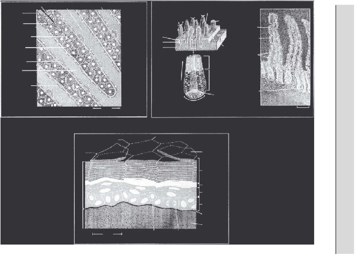
Liver
Intestinal Epithelium
LUMEN OF GUT
bile to gut
blood flow
epithelial cell migration
from “birth” at the bottom
of the crypt to loss at the
top of the villus
(transit time is
3-5 days)
bile duct
Villus (no cell division)
cross-section
of villus
fibroblast
villus
absorptive
brush-border
cells
epithelial
cells
bile canaliculus
crypt
cross-section
of crypt
hepatocyte
loose
connective
tissue
mucus-
secreting
goblet cells
nondividing
differentiated
cells
rapidly dividing
cells (cycle time =
11 hours)
Kupffer cell
direction of cell
movement
sinusoid
endothelial cell
slowly dividing stem
cells (cycle time >
24 hours)
crypt
nondividing
differentiated
cells
(b)
50
μ
m
m
From The Art of MBoC
3
© 1995 Garland Publishing, Inc.
100
μ
(b)
(a)
From The Art of MBoC
3
© 1995 Garland Publishing, Inc.
Skin
squame about
to flake off
from surface
keratinized
squames
granular
cell layer
prickle
cell layers
basal
cell layer
basal
lamine
connective
tissue of
dermis
basal cell
passing into
prickle cell layer
basal cell dividing
30
μ
m
FIGURE 6.25
In vivo tissue microenvironments.
From Alberts et al., 1995.