Biomedical Engineering Reference
In-Depth Information
4
3.5
Control Ventricle
3
2.5
2
Decreased Contractile State
1.5
1
0.5
0
0
0.25
0.5
0.75
1
Time [s]
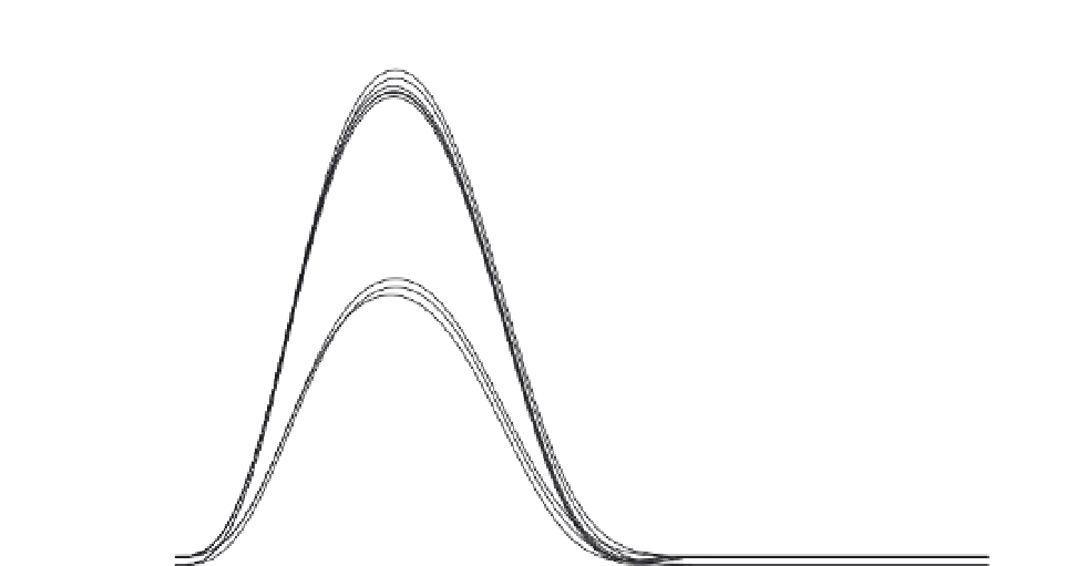
FIGURE 4.45
Ventricular elastance curves computed using the new analytical function of Eq. (4.77). Elastance
curves computed in this way are representative of the ventricle's contractile state—that is, its ability to pump blood.
invariant and cluster in two groups: either normal or weakened ventricle contractile state.
Consequently, this new measure of elastance may now effectively assess the health of the
heart alone, separate from blood vessel pathology.
Chapter 3 gives a brief overview of the circulatory system, a mass and heat transfer
system that circulates blood throughout the body. Figure 3.18 shows the four chambers of
the heart, the major blood vessels, and valves between the two. From a mechanical point
of view, the contracting heart chambers generate pressures that propel blood into the down-
stream blood vessels. This process is depicted in detail for the most important chamber, the
left ventricle, in Figure 4.46. Plotted are representative waveforms for an isolated canine
left ventricle, with ventricular pressure and root aortic pressure (top), ventricular volume
(middle) and ventricular outflow (bottom), all as functions of time. The numbers 1-4 at
the top of the figure correspond to four major phases of the contraction cycle, marked by
dashed lines. At time 1, filling is complete, the mitral valve closes, and the ventricle begins
to contract isovolumically (no change in volume). At time 2, ventricular pressure exceeds
root aortic pressure, the aortic valve opens, and blood ejection begins. Heart valves are
passive, and the outflow of blood results simply from the pressure difference across the
valve. When ventricular pressure falls below aortic pressure, the aortic valve closes (time
3) and outflow ends. The initial volume (time 1) is denoted end-diastolic volume, EDV,
and the volume at the end of ventricular ejection (time 3) is end-systolic volume, ESV.
At time 4, ventricular pressure falls below left atrial pressure (not shown), the mitral valve
opens, and filling begins in preparation for the next heartbeat. The difference between


