Biomedical Engineering Reference
In-Depth Information
200
300
180
250
160
Ved = 45ml
SV = 23ml
EF = 50%
140
200
120
150
100
80
100
60
40
50
20
0
0
0
0.25
0.5
0.75
1
Time [s]
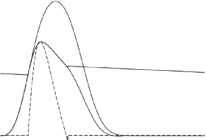
FIGURE 4.44
The same normal canine ventricle of Figure 4.43 now pumping into an arterial system with
doubled peripheral (flow) resistance. As expected, increased resistance, corresponding to narrowed vessels, leads
to increased arterial pulse pressure. Stroke volume is reduced from 66 to to 50 percent.
As expected, increased peripheral resistance raises arterial blood pressure to 140/95 mmHg
and impedes the ventricle's ability to eject blood (Figure 4.44). The ejection fraction decreases
to 50 percent in this experiment. Other experiments, such as altered arterial stiffness, may be
performed. The model's flexibility allows description of heart pathology as well as changes in
blood vessels. This one ventricular equation with one set of measured parameters is able to
describe the wide range of hemodynamics observed experimentally [24, 23].
The previous expressions for ventricular elastance defined in Eqs. (4.73) and (4.74) have
the same units as elastance defined classically as Eq. (4.72), but are mathematically not the
same. Since ventricular pressure is defined as an analytical function (Eq. (4.75)), ventricular
elastance,
E
v
, defined in the classical sense, may now be calculated as
@
p
v
=@
V
v
:
"
#
Þ
e
ð
t
t
b
a
a
e
ð
t
c
Þ
r
Þ
ð
1
t
E
v
ð
t
,
V
v
Þ¼
2
a
ð
V
v
b
Þþ
c
ð
4
:
77
Þ
e
ð
t
t
c
Þ
Þ
e
ð
tp
t
b
a
a
r
Þ
ð
1
t
or
E
v
ð
t
,
V
v
Þ¼
2
a
ð
V
v
b
Þþ
cf
ð
t
Þ
ð
4
:
78
Þ
Figure 4.45 shows ventricular elastance curves computed using this new analytical defi-
nition of elastance (Eq. (4.77)). Elastance was computed for a wide range of ventricular
and arterial states, including normal and pathological ventricles, normal and pathological
arterial systems, and isovolumic and ejecting beats. These elastance curves are relatively


