Biomedical Engineering Reference
In-Depth Information
(A)
(a)
(b)
(B)
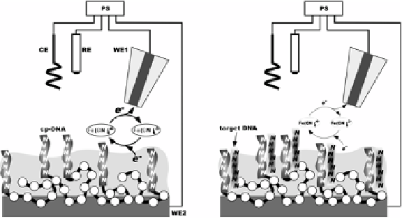
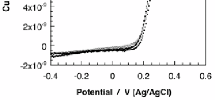
Figure 12. (A) Schematic illustration for the SECM imaging at the CP-DNA/PQ
microdots (
a
) and the surface state after hybridization (
b
). (B) Detection of hybrid-
ization of CP-DNA confined in the substrate electrode with the tip CV measure-
ments. CVs were obtained with a potential scan rate of 50 mV s
-1
and the substrate
was fixed at -0.2 V. During the measurements, the electrodes were hold adjacent at
20 μm-distance. The substrate surface was varied form the CP-DNA-attached (
a
) to
the hybridized state (
b
). The mediator solution was 10 mM Fe(CN)
6
4-
containing
0.1 M KCl.

Search WWH ::

Custom Search