Biomedical Engineering Reference
In-Depth Information
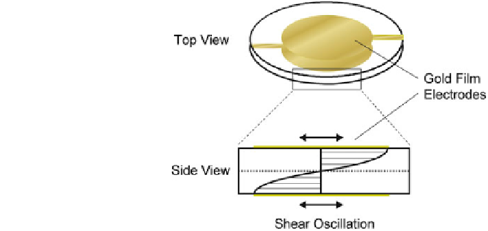
Fig. 13 Top and side views
of a shear wave resonator as
used in QCM-based
experiments. The quartz
resonator is sandwiched
between two gold film
electrodes used to drive the
resonant oscillation and to
read the resonance frequency.
Under resonance conditions a
standing acoustic wave is
established between the
crystal's surfaces. Resonance
parameters are very sensitive
to adsorption or desorption
processes at the surface
determine the time T
1/2
needed to attain half-maximal capacitance decrease after
wounding and, thus, half-maximal repopulation of the electrode (= wound healing).
The slope of the capacitance versus time curves mirrors the migration velocity.
Fluorescence microscopic observation of the ECIS-based wound healing assay
(Fig.
12
b) provides images of the different stages of the wound healing process.
A fluorescence-based viability assay based on ethidium homodimer-1 (EthD-1; red
fluorescence) and calcein acetoxymethylester (CaAM; green fluorescence) was used
to discriminate live and dead cells in the micrographs at the times indicated in
Fig.
12
a. The DNA-intercalating dye EthD-1 is a marker for membrane integrity as it
is non-membrane-permeable and can only access the nuclei after membrane
permeabilization. Calcein AM (CaAM) is essentially non-fluorescent but
membrane-permeable. Intracellular esterases inside living cells hydrolyze CaAM to
the membrane-impermeable calcein, which emits a green fluorescence. Before the
high-field application, all cells exhibit a green cytoplasmic fluorescence, which thus
indicates vital cells. After the elevated field is applied, all cells residing on the
electrode are selectively wounded as indicated by their EthD-1 stained cell nuclei
(red), while the cells surrounding the electrode remain vital, showing a green cyto-
plasmic fluorescence (Fig.
12
b2). Figure
12
b3 shows a fluorescence image of an
electrode covered with NRK cells after half-maximal wound healing. A radial
growth pattern in the cell layer near the electrode periphery can be observed as the
cells have migrated inward, suggesting a re-alignment of the cells during the
migration process. This pattern is even more pronounced for the image recorded after
wound closure (Fig.
12
b4).
4.4 Acoustic Techniques for Studying Cell-Surface Interactions
Several acoustic approaches have been described that are capable of providing
valuable information about the formation and modulation of cell-surface inter-
actions. By far the most widely known device is the quartz crystal microbalance
Search WWH ::

Custom Search