Biomedical Engineering Reference
In-Depth Information
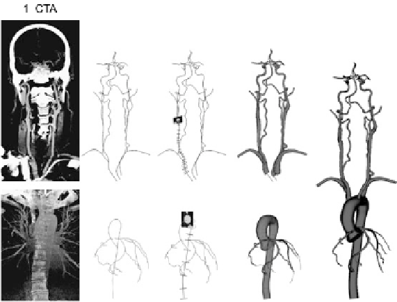
Fig. 15.1
3D computational model of the human thoracic aorta, coronary arteries, and head and
neck vessels based on CT images from two normal male subjects. From Coogan et al. (
2012
)
code SimVascular (Figueroa et al.,
2006
). Computations were performed on a super-
computer (276 Dell PowerEdge 1950) typically using 96 cores. We utilized a time
step size of 0.0001 seconds, and the simulations had an average residual of 0
.
005.
Simulations were run for 7 cardiac cycles until achieving cycle-to-cycle periodicity
in the pressure fields.
15.2.3 Fluid-Solid Models
1
.
06 kg/m
3
Blood density was
ρ
0
.
04 P. We as-
sumed typical baseline values for the linearized stiffness and thickness of the wall
of each of the four primary vascular segments: thoracic aorta as well as coro-
nary, neck, and cerebral arteries. A coupled momentum method was used (Figueroa
et al.,
2006
) to model wall deformability and a coupled-multidomain formulation
(Vignon-Clementel et al.,
2006
) was used to link Windkessel models for the heart
and distal vessels to the 3D vascular model. The overall model thus required pre-
scription of one inlet and 22 outlet boundary conditions. Numerical values for the
lumped-parameter coefficients were determined iteratively to reach target values for
flow and pressure.
=
and blood viscosity was
μ
=