Biomedical Engineering Reference
In-Depth Information
(a)
(b)
Gd
2
O(CO
3
)
2
T
1
shell (1.5 nm)
SiO
2
separating
layer
:
T
2
contrast
materials
:
T
1
contrast
materials
MnFe
2
O
4
T
2
core (15 nm)
20 nm
(c)
(e)
40
2
3
1
T
1
image
30
20
1: Feridex
2: Gd-DTPA
3: DMCA
10
Gd-DTPA
DMCA
0
0
5 0
15
20
1
2
(d)
500
400
3
T
2
image
MnFe
2
O
4
300
200
Feridex
100
0
0
5 0
SiO
2
thickness (nm)
15
20
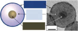
fIgure 2.10
(a) Schematic illustration of the magnetic coupling of a
T
1
contrast agent and
a
T
2
contrast agent in proximity. (b) Structure and HRTEM image of “magnetic-decoupled”
dMca. (c) Plot of relaxivity
r
1
value and (d) relaxivity
r
2
value
versus
the thickness of
separating SiO
2
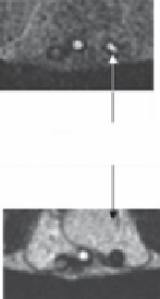
layer in dMca. (e)
In vivo T
1
-weighted MR image and
T
2
-weighted MR image
of mice implanted by dMca, Ferridex, and gd-dTPa in separated tubing. (Reprinted with
permission from Ref. [56]. © american chemical Society.)
the sensitivity and efficiency of biomedical imaging. The high magnetization Fe NPs
with Fe
3
O
4
and ferrite protective shell provided another appealing class of probe for
sensitive MRI. To optimize the efficiency of biomedical imaging, IOMNPs have
been synthetically combined with other NP components in different hybrid formats
such as core-shell and dumbbell, enabling superior multimodality imaging. The
as-fabricated MRI-optical, MRI-cT, and MRI
T
1
-
T
2
multimodality nanoprobes
have significantly enhanced the biological imaging in accuracy, sensitivity, and
special/time resolution.
despite the exciting progress in this field, the IOMNPs are still in lab demon-
stration stage. The advanced MRI and multimodality imaging require the
formation of IOMNPs with high magnetic moments, high stability, and biocom-
patibility, which should be further improved. The surface engineering of IOMNPs


Search WWH ::

Custom Search