Biomedical Engineering Reference
In-Depth Information
in the same liposome display differential release profile
in vivo
[105]. This example
illustrates the need for careful selection of imaging agent and the drug in a theranos-
tic formulation, and how the drug and imaging agent are incorporated will drive the
theranostic performance.
15.3.2
nanoemulsions
nanoemulsions are kinetically stable emulsions with a droplet size typically between
100 and 500 nm, high oil content, and low amounts of surfactant [106]. nanoemulsions
have wide applications in pharmaceutical and food industry. nanoemulsions are
typically used to increase tissue penetration of poorly soluble drugs (transdermal),
drug solubility, and bioavailability (injectable) [107-111]. nanoemulsions are also
easily incorporated into other dosage forms such as capsules and gels. They can be
produced on an industrial scale [111-113]. Therefore, due to scalability and high
drug carrying capacity, nanoemulsions also represent an attractive theranostic
development platform with a high potential for clinical use.
in nanoemulsions, imaging modalities that have been explored are Mri, ultra-
sound, photoacoustic, and optical imaging. perfluorocarbons (pFCs) are so far the
most utilized materials for imaging nanoemulsions. pFCs for Mri
in vivo
were first
reported by longmaid
et al
. [114]. They used pFC nanoemulsions, originally
designed as blood substitutes, and imaged the liver, tumor, and abscess in rats. since
this report, pFC nanoemulsions were explored extensively as Mri agents [115].
recently, ahrens and Zhong summarized the use of pFCs for cell tracking
in vivo
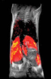
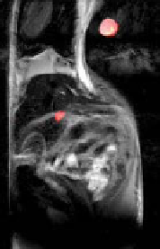
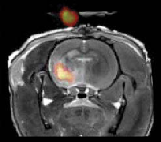
[116]. Figure 15.4 shows typical examples of
19
F Mri using pFC nanoemulsions.
(a)
(b)
(c)
R
Lu
P
L
L
S
figure 15.4
In vivo
19
F Mri using
ex vivo
perfluorocarbon (pFC)-labeled cells in rodent
models.
19
F images of the labeled cells are displayed on a “hot-iron” intensity scale, and the
anatomical (1h) images are shown in gray scale. (a) Composite image through the torso follow-
ing intravenous inoculation with labeled dendritic cells (DCs). Cells are apparent in the liver
(l) and spleen (s) and weakly in the lungs (lu). (b) adoptively transferred T cells selectively
home to the pancreas in a prediabetic mouse model. The image shows T cells (pseudocolor)
homing to the pancreas (p). r, reference capillary. (c) neuronal stem cells injected into the
infarct of a rat stroke model. a
19
F reference capillary is outside the brain. (reprinted from ref.
[116]. © John Wiley & sons, ltd.) (
See insert for color representation of the figure.)
.)
Search WWH ::

Custom Search