Biomedical Engineering Reference
In-Depth Information
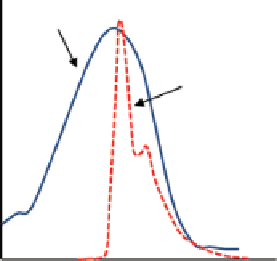
Absorption spectrum
of quencher
Emission spectrum
of uorophore
Wavelength (λ)
FIgure 9.3
Dynamic quenching occurs when the absorption spectrum of a quencher over-
laps the emission spectrum of a fluorophore. To generate photons at the emission wavelength
of a quencher, the distance between the fluorophore's and the quencher's molecules should be
smaller than 100Å.
Static quenching happens when a complex formation in ground state of a fluoro-
phore and a quencher occurs. In this case, the complex is a completely new material
with its own unique properties and excitation and emission spectra that are different
from the original fluorophore's and quencher's spectra.
Dynamic quenching is the basis for fluorescence resonance energy transfer
(FreT) assays and fluorescent probes that can turn on in specific conditions and
turn off in others. The fluorophore and quencher are usually referred to as donor
and acceptor molecules. Dynamic quenching is the basis for activatable optical
contrast agents for molecular imaging [25, 29]. using activatable fluorescent
probes that can turn on and turn off can improve the image quality and signal-to-
background ratio.
The parameters that have to be considered in designing a fluorescent probe for
biomedical applications, in addition to its optical properties, are stability, toxicity,
and molecular size. The molecular size of a fluorescent probe is important since it
determines the pharmacokinetics and clearance time of the probe from the blood
circulation and normal tissues.
Biological tissues contain many endogenous fluorophores such as amino acids,
lipofuscins, melanin, collagen, elastin, nicotinamide adenine dinucleotide (NADH),
and flavins [30, 31]. These fluorophores mainly fluoresce in ultraviolet or visible
wavelengths. In near-infrared light, the autofluorescence of the tissue is negligible,
and in many
in vivo
systems, it is below the noise level. Therefore, using exogenous
fluorophores that emit in near-infrared spectrum has a benefit of no interference with
autofluorescence of tissue.
An alternative approach to fluorescence probes is phosphorescence probes [32,
33]. Phosphorescence probes have a broader temporal response (microsecond range)
compared to fluorescent probes (nanosecond range). Due to their longer temporal
Search WWH ::

Custom Search