Biomedical Engineering Reference
In-Depth Information
+
-
+
Ag
Au
Au
Au
-
Dye-NP
Free dye
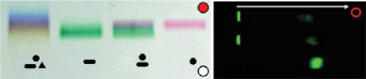
figure 6.25
left: separation of gold and silver nanoparticles according to their size and
shape by agarose gel electrophoresis after coating them with a charged polymer layer. (Reprinted
with permission from Ref. [103]. © american Chemical Society.) Right: gel electrophoresis of
NIR-labeled nanoparticles after synthesis before purifications, negatively charged free dye
migrates to the cathode, while labeled nanoparticles remain at the site of loading.
development includes the optimization of electrophoretic conditions through adjust-
ments of the potential between the electrodes, agarose/polyacrylamide density, and
buffers to achieve sufficient separation of an unreacted targeting molecule from the
nanoparticle. Relatively large nanoparticles lack mobility and retain at the loading
point of the gel, while smaller size targeting moieties such as peptides, proteins, and
antibodies migrate through the gel matrix toward one of the electrodes (fig. 6.25). The
gel bands can be then quantified by any optical imager if a fluorescent label is used or
by other means. Such methods have been used to monitor the synthesis and the
stability of encapsulated ICg and used to assess the purity of the nanoparticle [40].
6.8.2
chromatography
Chromatographic methods, where the separation is based on the differences in the
partition coefficients between mobile and stationary phases for all components of the
mixture, have long been used for nanoparticle purification. The method is occasion-
ally used to evaluate the size of the nanoparticles [104].
6.8.3
quartz crystal microbalances
This method provides a simple yet cost-effective and high-resolution mass sensing
technique based upon the piezoelectric effect [105] and is typically utilized for
measuring nanoparticle concentration [106]. The past two decades have witnessed an
explosive growth in the application of the quartz crystal microbalance (QCM) tech-
nique to the study of a wide range of molecular systems at the solution-surface inter-
face, in particular biopolymer and biochemical systems, but its full potential in
nanoparticle research has yet to be demonstrated.
6.8.4
flow cytometry
flow cytometry is essentially a single particle analysis, which provides rapid
information regarding the size, shape, complexity, as well as optical properties of
thousands of particles within several seconds. The principle of the method is based
on the interaction of particles with a beam of light. Three major components of these
interactions—forward scattering, side scattering, and fluorescence intensity (in the
Search WWH ::

Custom Search