Biomedical Engineering Reference
In-Depth Information
T
2
relaxation
100
75
T
2
= 150 ms
T
2
= 100 ms
Contrast (5×)
50
25
0
0
100
200
Time (ms)
300
400
500
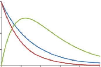
figure 6.16
MRI signal intensity (arbitrary units) for tissues with
T
2
values of 150 ms or
100 ms over a range of TE values. The tissue with the shorter
T
2
decays quicker than the tissue
with the longer
T
2
. Subtracting the MRI signals provides the image contrast between the two
tissues (expanded fivefold to aid visualization). The maximum image contrast occurs with a
TE of 120 ms.
are broadly separated into two separate categories,
T
1
or
T
2
agents.
T
1
agents usually
consist of a paramagnetic metal, while
T
2
agents consist of superparamagnetic iron
oxide nanoparticles. The most common paramagnetic contrast agent used in MRI is
gadolinium-diethylenetriamine pentaacetic acid (gd-DTPa) [5-7]. This agent
decreases the relaxation times of nearby hydrogen nuclei. In proton density images,
the contrast agent does not significantly alter the image. In
T
1
-weighted images, the
T
1
is reduced, and tissues exposed to the contrast agent appear brighter. In
T
2
-weighted
images, the contrast agent decreases the
T
2
of tissues containing contrast agent, and
so they appear darker. The additional contrast may be used to detect hemorrhage,
disruption of the blood-brain barrier, or the increased vascularity associated with
tumors.
6.6.2
relaxivity
The ability of a contrast agent to alter the MRI relaxation times is denoted by its
relaxivity [8]. Contrast agents alter the
T
1
relaxation time by
11
c
=+
[]
rP
1
T
T
1
1
where
T
1
c
is the relaxation time with the contrast agent,
T
1
is the relaxation time
without the contrast agent,
r
1
is the longitudinal relaxivity, and [
P
] is the contrast
agent concentration. The
T
2
relaxivity is similarly given by
11
c
=+
[]
rP
2
T
T
2
2


Search WWH ::

Custom Search