Biomedical Engineering Reference
In-Depth Information
Most of the contrast produced by samples on TEM is due to varying thickness and
density of the sample. Higher atomic number elements and thicker particles allow for
less transmission of electrons and therefore create a darker region in the image. In the
case of organic particles that are composed primarily of low atomic number ele-
ments, producing enough contrast to image the particles can be an issue. one way to
address low contrast in organic samples is to use a lower accelerating voltage.
another, more commonly used technique, is negative staining in which a sample is
treated with a solution containing a high atomic number element that will absorb
electrons (uranium, gold, lead, osmium). Successful negative staining depends on
the chemistry of the stain and sample interaction. a successfully stained sample
may appear either light with a dark background or dark with a light background
(an example is shown in figs. 6.5 and 6.6).
Particle sample preparation for TEM is usually a fairly simple matter involving
drop casting or dip coating of particles in a solution onto a standard carbon-coated
copper grid and allowing them to dry. for organic particles, drop casting or dip coat-
ing of particles is often followed by a negative staining procedure. for particles that
cannot tolerate drying, a cryogenic sample holder can be used to image particles
frozen in liquid nitrogen.
TEM is also used for imaging of intracellular features of cell and tissue samples. It
is sometimes possible to image particles present in cell and tissue samples; however,
such imaging requires extensive sample preparation including fixation, polymeriza-
tion, ultrathin sectioning, and negative staining of the sample. as most tissue and cell
samples are measured in microns or millimeters, locating nanoparticles can be a
time-consuming affair. likewise, this technique is not useful for particles that may be
difficult to distinguish from naturally occurring organelles and cellular features.
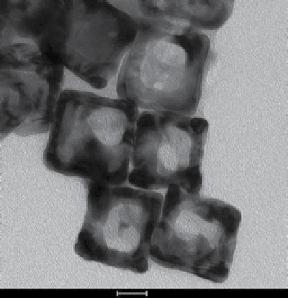
20 nm
figure 6.5
Transmission electron micrograph of gold “nanocages” drop cast on a TEM grid.

Search WWH ::

Custom Search