Biomedical Engineering Reference
In-Depth Information
6.3.1
tem
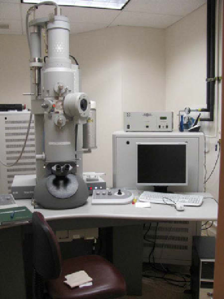
TEM is one of the most popular tools for characterizing nanoparticles as it can
provide high-resolution images of particles ranging in size from less than one
nanometer to hundreds of nanometers. In a TEM, electrons are fired from an elec-
tron beam at voltages ranging from 20 kV to about 120 kV toward an ultrathin
sample (hundreds of nanometers or less) within a vacuum chamber, the electrons
interact with the sample, and an image is formed from the electrons that are trans-
mitted through the sample and are focused onto an imaging plane located below the
sample holder. an example of a TEM instrument is shown in figure 6.4. In most
modern TEMs, the focused image is collected using a CCD camera. as this tech-
nique utilizes electrons that have been transmitted through a sample, the image
produced is a two-dimensional (2D) profile, which for many particle samples
allows for easy measurement of particle diameter or edge length. However, as TEM
is a high-resolution technique, the field of view is very limited, making collection
of statistically relevant numbers of particle sizes a time-consuming task. for this
reason, TEM is most often used to confirm morphology and sizing results obtained
via different methods.
figure 6.4
Tecnai Spirit transmission electron microscope from fEI Company.
Search WWH ::

Custom Search