Biomedical Engineering Reference
In-Depth Information
(a)
(b)
(c)
1.1
0.9
BV
BV
0.7
0.5
SLN
0.3
0.1
0
50 100 150
Time (min)
Injection time
200
250
5 mm
5 mm
0.5
1
1.5
2
2.5
0.4
0.8
1.2
1.6
2
(d)
(e)
(f )
SLN
SLN
SLN
5 mm
5 mm
5 mm
0.1
0.2
0.3
0.4
0.5
0.6
0.7
0.8
0.05
0.1
0.15
0.2
0.25
0.3
0.35
0.4
0.05
0.07
0.09
0.11
0.13
0.14
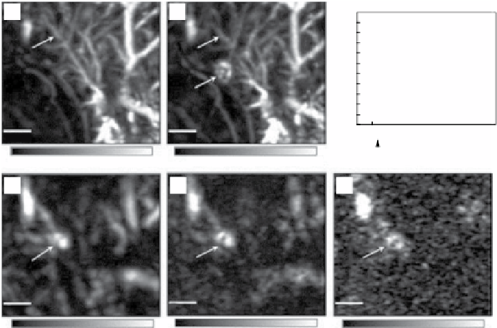
figure 5.16
OA images of the axillary region of a rat taken (a) before and (b) approxi-
mately 28 min after the injection of gold nanocages. (c) The changes of OA signal amplitude as
a function of the postinjection time. After the injection, OA signals increased with time, which
means gradual accumulations of the nanocages. (d-f) Depth capability of sLN mapping with
gold nanocages. The OA images were acquired after the injection of nanocages for (d) 126 min
with a total imaging depth of 10 mm by placing a layer of chicken breast tissue on the axillary
region, (e) 165 min with a total imaging depth of 21 mm by adding another layer of chicken
breast tissue, and (f) 226 min with a total imaging depth of 33 mm by using three layers of
chicken breast tissue. The bars represent the optical absorption. BV, blood vessel. sLN,
sentinel lymph node. (Reproduced with permission from Ref. [152]. © American chemical
society.)
A study [153] successfully demonstrated the use of gold nanocages as a contrast
agent for quantitative PA imaging of melanomas
in vivo
. An additional advantage
(which is common to other gold nanoparticles) is the gold nanocages' ability to
effectively transfer light energy into the heat destructive to many tumors. Thus, the
absorption of NIR light by the immunotargeted nanocages resulted in the selective
PT destruction of cancer cells
in vitro
[154].
5.4.5
hollow gold Nanospheres and Nanoshells
Another type of plasmonic nanoparticle often used as a contrast agent for OAT and
imaging is hollow gold nanospheres/nanoshells (HAuNs). OA imaging of living
mouse brain vasculature using HAuNs has been demonstrated by Lu
et al
. [155].
figure 5.17 shows noninvasive OA imaging of a mouse brain
in vivo
employing
Peg-HAuNs and NIR light at a wavelength of 800 nm. At higher magnification on


Search WWH ::

Custom Search