Biomedical Engineering Reference
In-Depth Information
40
Extinction
Extinction
15
Extinction
30
30
Absorption
Absorption
10
Absorption
20
20
Scattering
Scattering
Au Nanocage
Inner edge length = 50 nm
t
= 6 nm
Scattering
Au Nanorod
w=20 nm, L=66 nm
5
10
10
Silica Core Au Nanoshell
D
shell
= 50 nm,
t
= 3.2 nm
0
0
0
400
600
800
1000
400
600
800
1000
400
600
800
1000
r
eff
= 8.74 nm
r
eff
= 11.43 nm
r
eff
= 17.90 nm
16
12
18
14
10
15
12
8
12
10
8
9
6
6
6
4
4
3
2
2
0
0
0
700
800
Wavelength (nm)
900
1000
700
800
Wavelength (nm)
900
1000
700
800
Wavelength (nm)
900
1000
R
= 3.1
R
= 3.9
R
= 4.6
14
16
18
12
14
15
10
12
12
8
10
8
9
6
6
6
4
4
3
2
2
0
0
0
700
800
Wavelength (nm)
900
1000
700
800
Wavelength (nm)
900
1000
700
800
Wavelength (nm)
900
1000
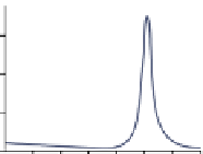
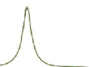
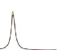
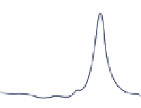
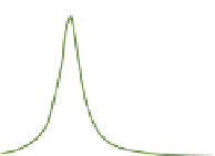
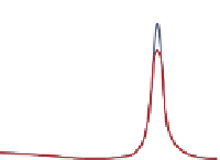
figure 5.11
upper Row: comparison of the optical spectra calculated using discrete dipole
approximation (DDA) for gold nanorods (gNR), nanocages (gNc), and nanoshells (gNs). for
all the structures, the surface plasmon resonance peak was tuned to exactly 800 nm. The gNR
has dimensions 20 nm × 66 nm. The gNc has an inner edge length of 50 nm and wall thickness
of 6 nm. The gNs has a silica core of 50 nm in diameter and shell thickness of 3.2 nm.
(Reproduced with permission from Ref. [27]. © John Wiley & sons, Inc.) (middle and
Bottom Rows): calculated spectra of the optical absorption efficiency,
Q
abs
(
light gray
),
scattering
Q
sca
(
black
), and extinction Q
ext
(
dark gray
) for gNR with fixed aspect ratio (R) = 3.9
and different effective radius (
r
eff
) 8.74, 11.43, and 17.90 nm (
middle
) , and with fixed
r
eff
= 11.43 nm
and different (R) 3.1, 3.9, and 4.6 (bottom). (Reproduced from [111] .with permission from
American chemical society).
5.4.2
gold Nanoshells
Nanoshells for biomedical applications usually consist of a dielectric core coated by
a conductive, nanometer-thick metallic shell, typically gold [112]. core size and/or
shell thickness can shift the sPR that allows nanoparticle optical properties to be
tuned over the visible and NIR spectral regions [113, 114]. Nanoshells are optically
robust, exhibiting resistance photobleaching and chemical/thermal stability to dena-
turation [115]. Nanoshells comprised of gold offer a straightforward means to chem-
ically modify surface characteristics by grafting on functional groups ranging from
Pegs to integrins [27, 116]. gold nanoshells were first adopted for OA imaging
around a decade ago [117, 118]. The initial application of nanoshells as an OA
contrast agent was to increase the NIR contrast of cortical blood vessels in rat brain


Search WWH ::

Custom Search