Biomedical Engineering Reference
In-Depth Information
Plain SWNT
SWNT-RGD
Tumor
photograph
5 mm
5 mm
Tumor
Tumor
Ultrasound
3 mm
3 mm
100
Photoacoustic
preinjection
Photoacoustic
4 h postinjection
Subtraction
image
0
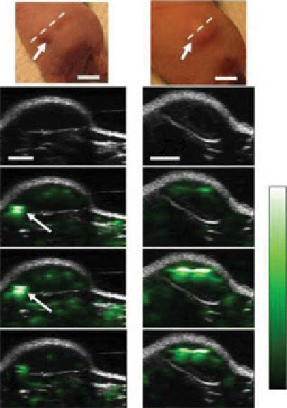
figure 5.8
Tumor targeting in living mice with nanoparticle conjugates of single‐wall
carbon nanotube and arginine-glycine-aspartic acid (sWNT‐RgD). The dotted lines on the
images illustrate the edges of each inclusion. A gray scale ultrasound B-mode image showing
the skin level was overlaid with the OA image (
white
). The high optoacoustic signal in the mouse
injected with plain sWNTs (white arrows) is not seen in the subtraction image, suggesting that
it is due to a large blood vessel and not sWNT. (Reproduced with permission from Ref. [87]. ©
Nature Publishing group.)
concentration of sWNTs was likely less than that because the PA images were
acquired before and after the administration of the contrast agent, which makes it
possible to separate the signal of the sWNT from the background.
Targeted sWNTs can be used for both OAT imaging and PA therapy. Recently, the
large OA effect of sWNTs was explored for targeting and selective destruction of
cancer cells [92]. under the irradiation of a 1064 nm Q-switched millisecond pulsed
laser, sWNTs showed a large PA effect in suspension, which could trigger an
explosion at the nanoscale. By conjugating the sWNTs with folic acid, which can
bind to cancer cells overexpressing folate receptors on the cell membrane, the laser
power used for cancer cell killing could be reduced 150-1500 times. This discovery
has new perspectives for exploring the PA properties of sWNTs in cancer therapy.
de la Zerda
et al
. summarized the vast applications of advanced nanoparticles with a
focus on cNT contrast agents for OA imaging, cytometry, and theranostics applications
Search WWH ::

Custom Search