Biomedical Engineering Reference
In-Depth Information
(a)
(b)
OH
-
H
+
1
2
3
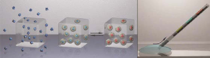
Figure 4.18
(a) The underlying concept of the sensor platform based on dye-loaded NCs
embedded in a highly permeable matrix: (1) low-molecular-weight dye molecules leach out
from the PVA matrix; (2 and 3) the same dye molecules encapsulated in porous NCs remain
entrapped in the PVA matrix. The porous nanothin wall of the capsules allows unhindered
transport of H
+
and OH
-
ions into the capsules. (b) A multiband capillary can sample a small
amount of fluid. (Adapted with permission from Ref. [65]. © American Chemical Society.)
An alternative imaging modality that can be used to image polymer vesicles is
diagnostic ultrasound. Zhou
et al
. prepared air-encapsulated polymersomes via
lyophilization and rehydration of previously formed polymer vesicles [76]. The
polymer bubbles were imaged using a Pie medical Scanner 350 and were visualized
as bright spots, validating the acoustic activity of air-encapsulated polymersomes.
ghoroghchian
et al
. [77] recently showed how to apply simple diblock copolymer
system such as poly(ethylene oxide)-block-poly(ε-caprolactone) (PEO-
b
-PCL) and
poly(ethylene oxide)-block-poly(γ-methyl-ε-caprolactone) (PEO-
b
-PmCL) to create
polymersomes with special emissive properties. This can be achieved by incorpo-
rating porphyrin-based fluorophores within the polymeric membrane. The final
structure shows optical properties similar to quantum dots from the visible to the
infrared. In another work, fluorescent amphiphilic molecules have been incorporated
to polymersome membrane and delivered within cells in order to generate a revolu-
tionary noncytotoxic, nonimmunogenic cellular tracking system [78]. Polymersomes
loaded with such fluorophores are therefore promising candidates for use as
“nanoscale imaging agents” [79]. In addition to the incorporation of fluorophores
within polymersomes [77, 78], the potential of those carriers to the field of imaging
has been further exemplified by the encapsulation of magnetic resonance imaging
(mRI) contrast agents within porous PEO-PBD polymersomes, in research work car-
ried out by Cheng and Tsourkas [80].
Additionally to fluorescent markers, quantum dots embedded into the capsules
can potentially be used as ion probes and luminescence sensors. The effect of pH
conditions and various ions on the luminescence intensity of CdTe nanocrystals
capped by thioglycolic acid has been studied [81]. Luminescence of both bare CdTe
nanocrystals and CdTe nanocrystals embedded into polymer microcapsules was
found to be pH sensitive within the range of pH 4-6. Another imaging application of
PEO-PBD copolymers has been found by the maskos research group, which encap-
sulates quantum dots within polymersome membranes [82].
NCs loaded with imaging contrasts or stimuli-responsive molecules can be
embedded into a highly permeable matrix. This method can lead to devices for med-
ical diagnostics (Fig. 4.18) that may use a broad range of optical chemosensors, such
Search WWH ::

Custom Search