Biomedical Engineering Reference
In-Depth Information
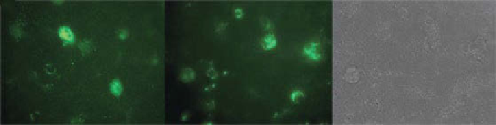
(a)
(b)
(c)
Figure 4.15
NCs (50 µg/ml) added to DRg neurons for 24 h, washed, and imaged.
(a) Single slice fluorescence only. (b) 3D slice fluorescence only. (c) 3D slice DIC only.
In the case of PmLCs, several research groups have assessed their toxicity by
performing
in vitro
cell viability assays such as the mTT test [68, 69]. generally, no
acute toxicity was observed at moderate capsule concentrations, and an outermost
polyanionic layer appeared to further decrease the toxicity (cationic PmLCs exhibit
a pronounced tendency to adhere to the cellular surface) [66]. Some toxicity was
observed at elevated capsule concentrations; this is commonly attributed to sedimen-
tation of the capsules on top of the cells as a result of competition for space between
the capsules and cells.
Picart
et al
. have shown that polyelectrolyte multilayers can be digested by enzy-
matic action when placed in the peritoneal [70] and oral environments [71]. The
biocompatibility and
in vivo
fate of dextran sulfate-poly-l-arginine polyelectrolyte
capsules after subcutaneous injection were recently assessed by De Koker
et al
. [68].
In our recent experiments, hollow polymer NCs loaded with calcein were taken up
by dorsal root ganglion (DRg) neurons (Fig. 4.15). Preliminary data indicate no
adverse effect of NCs on function of DRg neurons, consistent with data described
earlier.
In therapeutic applications, specific targeting of cells or receptors is desirable
because many side effects of current drugs emerge from the undesired impact of a
drug molecule on physiological pathways in a cell type or organ that is not involved
in the disease process [72, 73]. Targeting specifically functionalized polymersomes
to a desired cell type and location inside the body is of essential importance for
biomedical imaging (Fig. 4.16). Nanocontainer-treated macrophages showed
characteristic intracellular accumulations of vesicular structures with sizes in the
range of the nanocontainers. Together with the confocal microscopy results, this
finding is compatible with intracellular transport and accumulation of nanocontain-
ers to certain locations in the cells, presumably linked to the endocytotic pathway.
A variety of porphyrin-based or other near-infrared (NIR) dyes loaded to the
hydrophobic leaflet of polymersomes can be used for imaging
in vivo
. The total fluo-
rescence emanating from the polymersomes gave rise to a localized signal of
sufficient intensity to penetrate the dense tumor tissue of this living animal (Fig. 4.17),
giving a signal-to-background ratio of at least 10 to 1 [74]. Conjugation of Tat [75],
a cell permeable peptide, to NIR-emissive polymersomes allows tracking dendritic
cells. An improved and selective intracellular delivery of Tat-functionalized vesicles
was found in dendritic cells in comparison with nonfunctionalized polymersomes.
Search WWH ::

Custom Search