Biomedical Engineering Reference
In-Depth Information
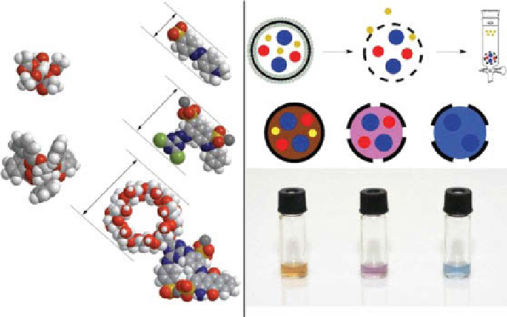
0.6 nm
(a)
(b)
1
1.1 nm
GPA
2
1.6 nm
GPB
3
Figure 4.8
Determination of pore size in vesicle-templated NCs using a selective perme-
ability assay. (a) Pore-forming templates. gPA, glucose pentaacetate, and gPB, glucose pen-
tabenzoate. Size probes: 1, methyl orange (0.6 nm); 2, Procion Red (1.1 nm); 3, Reactive Blue
2/b-cyclodextrin conjugate (1.6 nm). The smallest cross section corresponds to the smallest
pore the probe can cross in its most tightly packed conformation. (b) Selective permeability
demonstrated by the size probe retention assay. Liposomes were loaded with the mixture of
colored size probes: 0.6 nm yellow (1), 1.1 nm red (2), and 1.6 nm blue (3). monomers and
cross-linkers with or without pore-forming templates were loaded into the liposomal mem-
brane, and polymerization was initiated, after which the phospholipids and pore-forming tem-
plates were removed and capsules were separated from released size probes on a size-exclusion
column. The NC fraction is shown. In the absence of pore-forming templates, no probes
escaped, and the capsules remained brown (left sample). When gPA was used as pore-forming
template, 0.6 nm yellow probe was released, and the capsules were colored purple (middle
sample). With gPB as a pore-forming template, 0.6 nm yellow and 1.1 nm red probes were
released, and capsules were colored blue (right sample). (Reprinted with permission from Ref.
[14]. © Wiley.)
Functional groups in the pore orifice can be modified further using click chem-
istry [13]. For example, carboxylic groups in the nanopores can be activated and
converted to amide groups with a quantitative yield (Fig. 4.9). Functionalization of
nanopores did not compromise the ability to control the pore size (Fig. 4.9).
The NCs can retain entrapped molecules for extended periods of time. In a
previous study [17], we showed that polystyrene capsules did not release Procion
Red, a dye used as 1.1 nm size probe, for 240 days. A follow-up study on the same
samples showed no measurable release after 40 months. In these experiments, release
of as little as 1% of encapsulated dye could be detected with uV-Vis analysis of the
supernatant from the suspension of NCs. This long-term stability is likely the result
of high degree of cross-linking in the NC shells.
Search WWH ::

Custom Search