Biomedical Engineering Reference
In-Depth Information
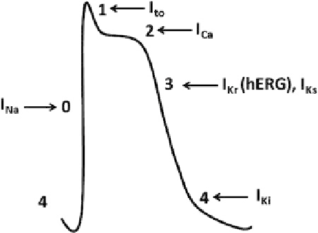
Fig. 2 Schematic diagram of an action potential recorded from a myocyte and involvement of Na
+
(I
Na
), K
+
(I
Kr
,I
Ks
, and I
Ki
) and Ca
2+
(I
Ca
) ion channel currents, which underlie each of its phases
(0, 1, 2, 3, and 4). The cardiac electric events are outcome of different biologically important ionic
pumps, exchangers and, most importantly, voltage-gated ion channels. The cardiac action poten-
tial recorded from either an atrial or a ventricular human myocyte can be classified into five
distinct phases. (a) The phase 0, phase 0, or action potential upstroke, is generated by the rapid,
transient influx of Na
+
into myocytes via Na
+
channels (inward current: I
Na
). (b) Phase 1 describes
the early repolarization process and is mediated by the efflux of K
+
from the cell through the
opening of a distinct K
+
channel. The transient outward current (I
to
) contributes to the termination
of the upstroke of the action potential by causing an early phase of rapid repolarization. (c) Phase
2, or plateau of the action potential, is essentially because of the entry of extracellular Ca
2+
into the
cells through L-type Ca
2+
channels (inward current: I
Ca
). (d) Phase 3 describes the late repolariza-
tion process and is mediated by the efflux of K
+
from the cell through the opening of distinct K
+
channels. Two distinct K
+
channels contribute to Phase 3 repolarization [I
Kr
or
hERG
current and
I
Ks
] by opposing Ca
2+
influx during the plateau phase. (e) Phase 4, resting potential, is maintained
by the inward rectifier (I
Ki
). Besides maintaining phase 4, the inward rectifier also plays a
prominent role in the final repolarization process in many species, although to a lesser extent in
the human heart and clamps the cardiac myocyte at its resting potential [
144
,
145
]
ventricular myocardium. Heterogeneity in development of prolongation of action
potential and early after depolarizations results in a myocardium that is vulnerable
to reentrant excitation, the probable immediate cause of
Tdp
in human [
43
].
2.1 QT Interval Correction (QTc)
The QT interval (Fig.
3
) and heart rate have an inverse, non-linear relationship,
which varies among and within species. Thus, change in heart rate exerts an effect
on QT interval, which can confound the assessment of effect of test substance on
ventricular repolarization and the QT interval [
44
]. Therefore, the interpretation of
data from
in vivo
test systems should take into account the effect of coincident
changes in heart rate. There are two important situations where variability in heart