Biomedical Engineering Reference
In-Depth Information
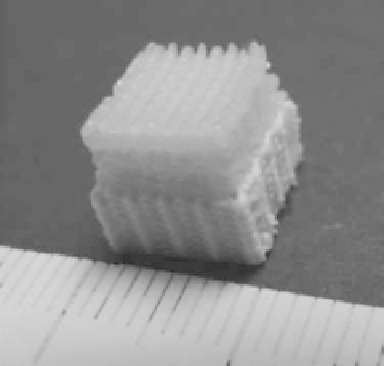
FIGURE 4.15
Photograph of a biphasic scaffold with separate layers for osteochondral tissue engineering.
The bottom layers were plotted from a CPC-alginate mixture for treatment of the bony part
and the top layers consist of alginate for the chondral part of the defect.
4.4 3D Plotting of Silicon-Based Bioceramic Scaffolds
Silicon-based bioceramics and scaffolds play an increasing role in bone tis-
sue engineering because silicon is one of the important trace elements in
human bone and is reported to stimulate new bone growth (Mertz 1981;
Pietak et al. 2007). Conventional methods for fabrication of porous silicon-
based bioceramic scaffolds are particle leaching and polyurethane template
sintering (Wu et al. 2010). As previously discussed, the drawbacks of these
methods do not only include poor control of pore parameters but also weak
mechanical properties of these scaffolds. Therefore, our intention was to
fabricate silicon-based scaffolds with designed pores with the application of
rapid prototyping.
In our work, two types of silicon-based scaffolds were printed by using
pastes containing either β-CaSiO
3
or mesoporous Ca-Si-P bioglass. β-CaSiO
3
is one of the most important silicon compounds used as bone replacement
material because β-CaSiO
3
degrades faster and has been demonstrated to
stimulate bone formation
in vivo
better than β-tricalcium phosphate (β-TCP)
(Xu et al. 2008). Ca-Si-P bioglass with regular nanochannels and high specific
surface area is a bioactive material. Numerous reports have demonstrated
that Ca-Si-P bioglass not only is an excellent drug delivery system but also
a potential candidate for bone repair (Lόpez-Noriega et al. 2006). β-CaSiO
3
powder was synthesized by a chemical precipitation method (Wu et al.
2012). Ca-Si-P bioglass was synthesized by a sol-gel and assembly technique
according to a published protocol (Wu et al. 2010). The product was ground

Search WWH ::

Custom Search