Biology Reference
In-Depth Information
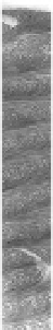
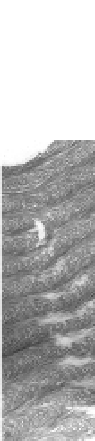
Figure 4.3
Transmission electron micrograph of a thin section cut through
an
silk moth chorion, showing the fibrous
ultrastructure of its lamellae. The parabolic pattern of fibres
(approximately 110 Å; dotted lines) within each lamella, in
oblique sections, is seen, which indicates that silkmoth chorion
is a biological analogue of a cholesteric liquid crystal.
11,12
Bar = 0.3 μm.
A. polyphemus
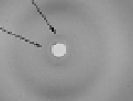
Figure 4.4
High angle X-ray diffraction pattern from an almost flat
fragment of a mature silkmoth chorion of
.
Incident beam is parallel to the chorion surface, which is
horizontal. The plane of the X-ray film is vertical. Note the
presence of 4.6, 9.1 and approximately 30 Å reflections,
indicative of a
A. polyphemus
β
-sheet structure for silk moth chorion proteins.
The 4.6 Å reflection corresponds to the repeat between
hydrogen bonded molecular polypeptide chains of chorion
proteins in a
β
-sheet conformation. The preferential orientation
of the 9.1 Å reflection indicates a preferential orientation
of stacked
β
-sheets parallel to the chorion surface, whereas
the elliptical scattering at approximately 30 Å indicates a
helicoidal architecture for silkmoth chorion and probably
arises from approximately 30 Å protofilaments, constituents
of the approximately 100 Å fibrils (Hamodrakas, 1992). A
A
toroidal camera was employed.


Search WWH ::

Custom Search