Biomedical Engineering Reference
In-Depth Information
90
µ
m
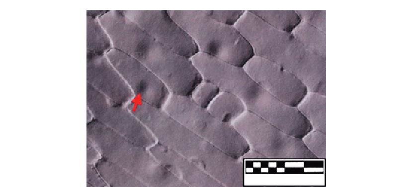
Figure 4.7
DIC image produced by software from data shown in
Figure 4.3
.
becomes uniformly gray. Upon placing the specimen on the stage of HMC microscope,
some of the incident light will be refracted and passed through either the 100% or the 1%
region of the modulator. Hence, the image of the specimen, as visualized by the slit, will
appear to be bright or dark, respectively. The incident light that is not refracted will still be
passing through the 15% region of the modulator. While the HMC microscope functions
like a DIC, it is immune to birefringence that can be caused by some specimens. Note that
the specimen must be rotated, allowing the asymmetries and symmetries in the specimen to
be differentiated. The structure of an HMC microscope and its operating concept is shown
in
Figure 4.8
.
It seems that this method has now lapsed as a patent and is still marketed under names such
as Varel by Zeiss
[17]
and by Nikon as NAMC
[18]
.
The author is unaware of its use for plant cells and tissue imaging thus far but it does have
strong potential in his field of endeavor.
4.7 Adaptive Optics Microscopy
Derived from astronomy primarily, it has so far only been used for fluorescence although it
should find use also in transmission and reflection imaging with quasicoherent light.
Recently, Zeiss has had precommercial instruments offering this technology.
A typical example of applying adaptive optics in the area of microscopy is demonstrated by
Poland et al.
[19]
. The experiment of this demonstration utilized a laser-scanning confocal
microscope (later reconfigured as a multiphoton fluorescent microscope) with DMM as its
Search WWH ::

Custom Search