Biomedical Engineering Reference
In-Depth Information
(A)
(B)
(C)
(D)
rad
8
6
4
2
0
(E)
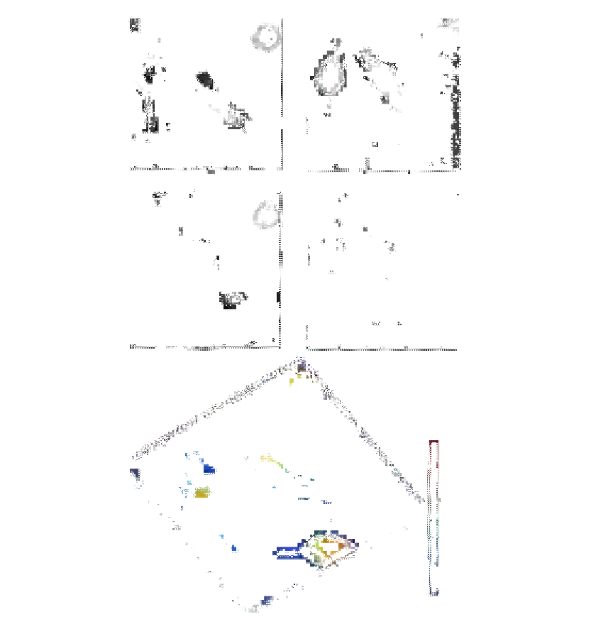
Figure 7.9
RAW cells—varying reconstruction distance unwrapping: (A)
(D) unwrapping stages and
(E) 3D rendering of the final unwrapped phase image. The images are 51.2
μ
m
51.2
μ
m
3
(256
256 pixels).
3
While this approach is superior to conventional phase unwrapping methods, it has its own
limitations. If the object is many wavelengths thick and/or the phase signal is very noisy,
that is, the phase images for different reconstruction planes are inconsistent, the method
begins to fail.
Figure 7.10
shows holographic image and the reconstruction of onion cells.

Search WWH ::

Custom Search