Information Technology Reference
In-Depth Information
10
6
1.0
0.9
0.8
10
5
0.7
0.6
0.5
10
4
0.4
0.3
0.2
10
3
0.1
0.0
0
0
6
6
12
12
18
18
24
24
30
30
36
36
42
42
48
48
54
54
60
60
66
66
72
72
time post-harvest (hour)
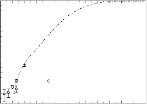
Fig. 5.
Simulation results using the parameter set presented in Table 1. The lines
represent the fraction of epithelial cells that are healthy (solid black), containing the
virus (dashed grey), secreting the virus (dashed black), or dead (dotted black), as well
as the number of competent virions (or pfu) on the right y-axis (dash-dot-dot black).
The diamonds and the circles represent experimental data for the viral titer and the
fraction of cells infected, respectively.
Note added in press
: Recent experiments have revealed a highly variable dynamic range
of the replication rate, but the basic structure of the model remains intact.
5
Proposed Extensions
As mentioned earlier, the current model is extremely simple, and we plan to
gradually increase the level of detail.
One of the first improvements would be the inclusion of different cell types.
The epithelial cells that make up the simulation grid are assumed to be a homo-
geneous population of cells, with no distinction, for example, between ciliated
and Clara cells. We plan to add more cell types; each cell type would have the
same four states illustrated in Fig. 1, and the transitions between those states
would still be dictated by the same processes, but the value of the parameters
controlling these processes would differ from one cell type to another and from
one virus strain to another. With such a model, we could, for example, explore
differences in the spread of the infection on a sample constituted of 90% cilliated
cells and 10% Clara cells against the spread on a sample constituted of 50%
ciliated cells and 50% Clara cells.
We also plan to break existing parameters into sub-models. Let us illustrate
this process with an example. At the moment, we describe viral release using the
parameter
g
V
which describes the constant rate at which virions are released by
secreting cells. In the future, this simple model of viral release could be replaced
by a much more elaborate intracellular sub-model of viral assembly and release
that takes account of factors such as viral strain and cell type to more accurately