Biology Reference
In-Depth Information
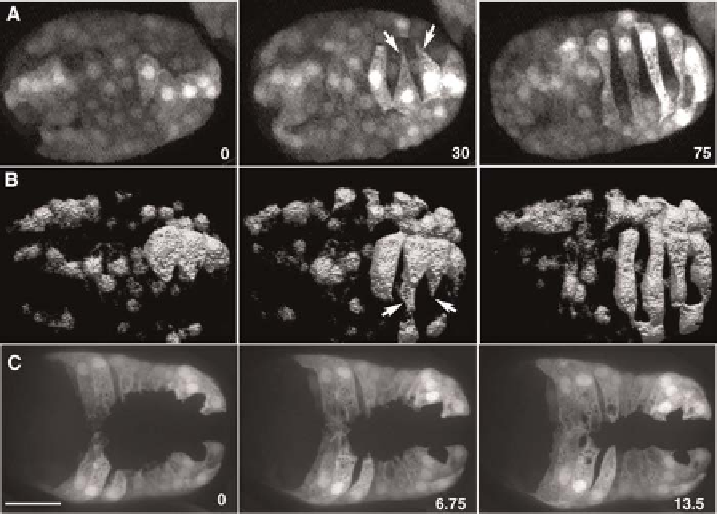
Fig. 6
Cytosolic markers allow imaging of cell motility and cell movement in embryos. (A, B) Frames
from 4Dmovies of dorsal intercalation in embryos expressing lbp-1p::gfp, which is expressed in a subset of
dorsal epidermal cells. (A) An embryo imaged using two-photon excitation microscopy. z-stacks were
subsequently projected using a maximum intensity procedure. (From Heid et al., 2001). (B) A similar
embryo imaged using a Perkin-Elmer UltraView LCI system (with Yokagawa CSU10 scanhead; images
courtesy of T. Walston). The dataset was subsequently subjected to surface rendering using Volocity
software. Fine protrusions are visible in both cases. It is clear in (B) that the protrusions are wedge-shaped
in the z-dimension. (C) Frames from a 4Dmovie of ventral enclosure in an embryo expressing a Pdlg-1::gfp
reporter. Elapsed time in minutes is shown. z-stacks were acquired at 50 s intervals, 20 focal planes/stack;
acquisition time/image, 300 ms. Fine details of protrusions are visible against a dark background using this
particular transcriptional reporter. (Images courtesy of M. Sheffield.) Bar = 10
m
m.
dramatically improving the effective contrast of the specimen being imaged.
Third, imaging cytosolic GFP reporters typically does not cause as much
photodamage as with GFP translational fusions. We have frequently used two
different markers to image events in the epidermis: Plbp-1::gfp (
Heid et al.,
2001
) and Pdlg-1::gfp (
Sheffield et al., 2007
). An example of the former is
shown in
Fig. 6A-B
, and the latter in
Fig. 6C
. An extremely useful adjunct to
the use of cytosolic markers is the use of a voxel rendering program, such as
Volocity (Perkin-Elmer).
Fig. 6B
shows the results this procedure in the case
of intercalation of dorsal epidermal cells. The result is a striking 3D view of
cells as they intercalate.
Search WWH ::

Custom Search