Chemistry Reference
In-Depth Information
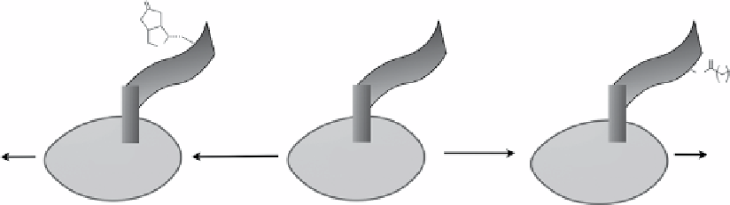
Peptide substrate
for biotin or lipoic acid ligase
O
S
O
N
3
N
7
H
Biotin ligase
substrate ketone
Lipoic acid ligase
substrate azide
Target protein
in cell
Oxime/hydrozone
ligation
Target protein
in cell
Staudinger ligation
azide-alkyne cyclisation
Target protein
in cell
fIgure 2.8
Protein labelling using biotin ligase or lipoic acid ligase peptide substrate tags.
demonstrated the accessibility of the azido group using its reaction with a cyclooctyne-conjugated fluorescent probe, and the
azido-modified cell surface fusion protein was visualised in live cells. The orthogonal use of LplA and biotin ligase was also
verified. one interesting application of this LplA-mediated labelling was illustrated in a study of protein-protein interaction
[319]. A photo-crosslinker, aryl azide, was introduced to one protein fused to LplA's peptide substrate, and the interacting
protein can be cross-linked upon uV irradiation.
Sortase Peptide Substrate Tag
Sortase, a class of bacterial transpeptidase, is in charge of anchoring cell surface proteins to
the cell wall by catalysing the cleavage of the threonine-glycine bond on the conserved LPXTg peptide substrate of its target
protein, followed by formation of a new bond between threonine and amino group on the cell wall. Mao et al. first demon-
strated the utilities of sortase in introduction of proteins, peptides, and small molecules to protein with LPXTg motif [320].
Since then, sortase-mediated ligation has been applied in protein labelling with functional ligands, protein imaging in living
cells [321, 322], protein immobilisation, protein purification, and construction of neoglycoconjugates [323, 324].
2.3.5.3 Introduction of Protein Tags
scFV tag
during the early time of developing surrogates to fluorescent proteins, the high affinity and specificity of the
antibody-hapten interaction has been applied in protein labelling strategies. Single-chain antibodies (scFV) targeting speci-
fied sites in living cells could arrest cell-permeable hapten-fluorophore conjugates through their highly affinity interactions
(nanomolar range) and were able to image specific subcellular localisation expressing the scFV, such as the endoplasmic
reticulum, golgi, and plasma membrane of living mammalian cells [325]. Chemical probes with various spectral and
indicator properties could be used in this approach.
FKBP12:F36V Tag
nolan and co-workers developed a protein labelling approach using high-affinity interaction between
an FKbP12 mutant (F36V) and a synthetic, engineered ligand SLF′ [326]. A protein fused with FKbP12:F36V tag could be
labelled noncovalently by SLF′-fluorophore conjugates in live mammalian cells, and the level of staining was proportional
to the expression level of the fusion protein. After SLF′-fluorophore labelling, β-galactosidase-FKbP12:F36V fusion protein
lost its enzymatic activity by 90% when fluorophore assisted laser inactivation was applied.
SNAP-tag or hAGT Tag
Human o
6
-alkylguanine-dnA-alkyltransferase (hAgT) is a 20 kda dnA repair protein that deal-
kylates o6-alkylguanine in damaged dnA. 'SnAP-tag' uses o
6
-guanine-modified fluorophore to covalently label proteins in
living cells (Scheme 2.25) [327]. This represents the first example using fusion protein technology to label target proteins
covalently and has been applied successfully in protein labelling in live mammalian cells, super-resolution imaging of intra-
cellular proteins [328], cell surface protein labelling for protein-protein interaction studies and virus-cell interaction studies
[329, 330], two-step protein immobilisation on solid surface [331], and quantum dots modification [332].
To image the reactive oxygen species in cells, SnAP-tag fusion proteins on the surface or interior of living cells were
labelled with boronate-capped dyes that could turn on in response to changes in local peroxide species levels [333]. A
tumour-targeting anti-egFR antibody fused to the SnAP-tag allowed specific attachment of o
6
-guanine-modified photosen-
sitiser at the target sites, increasing the phototoxicity in tumour cells [334]. Similarly, CLIP-tag, an AgT variant using o
2
-
benzylcytosine derivatives as substrate, has also been developed as an orthogonal analogue of SnAP-tag [335]. SnAP and
CLIP fusion proteins can be labelled with different imaging probes specifically and simultaneously in living cells.
HaloTag
bacterial haloalkane dehalogenases can replace halides from aliphatic hydrocarbons by nucleophilic attack from
aspartate in the enzyme to form a covalent ester bond with the hydrocarbon substrate (Scheme 2.26) [336]. Protein fused