Chemistry Reference
In-Depth Information
The isolated nanochains were found to consist of three to five particles. After cleavage from the beads, the mixture was
found to be 6% monomeric SPIONs, 72% four SPIONs, 12% three SPIONs, and 10% five SPIONs. The relaxivity of the
nanochains was found to be 121 mM
-1
s
-1
at 1.4 T, which is approximately twice that of the individual nanoparticles. Vivotag
680 was included for fluorescence imaging.
These nanochains were further modified with cyclic RGD to target a
v
B
3
integrin. The cyclic (Arg-GlyAsp-D-Phe-Cys)
was attached to the nanochains via maleimide chemistry. Nanochains with cRGD targeted tumours
in vivo
more efficiently
than either non-targeted nanochains or targeted nanoparticles, most likely due to the multivalent effect of the chain
structure (Figure 16.27). Notably, MRI was used to detect the presence of the targeted nanochains in liver and lung
metastases.
16.6.3
Medical applications
Tantalum oxide nanoparticles were investigated for SLN mapping using fluorescence and CT [9]. A microemulsion was
prepared using Igepal CO-520 (Figure 16.28
)
and tantalumethoxide was added. Rhodamine B isothiocyanate (RITC) was
functionalised with a silane group by mixing with APTES. The RITC-silane and PEG-silane were added to the tantalum
microemulsion to modify the surface of the nanoparticle.
Cell studies using MTT showed high viability. Histology showed low toxicity of injected particles. For SLN mapping,
particles were injected in the paws of mice. 3D CT imaging showed the location of the SLNs, which were successfully
removed using fluorescence-guided surgery.
Atherosclerotic disease is typically investigated using angiography. In order to identify areas of probable rupture, there is
a need for the ability to image the vessel wall. In this study, PET and MRI were combined to address this need [8]. Dextran-
coated SPIONs with amine functional groups were modified with
p
-NCS-Bn-DOTA-
64
Cu (Figure 16.29). PET was used to
screen for inflamed lesions while macrophages were visualised using MRI at high resolution. This 'non-invasive' endoscopy
shows macrophage density and distribution, which can in turn be used to estimate the probability of rupture (Figure 16.30).
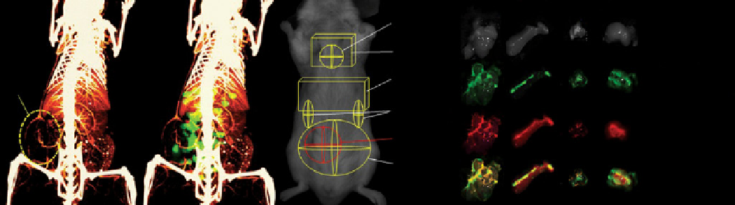
(a)
(b)
(c)
(d)
Liver
Spleen
Lungs
Tumour
Heart
Bright
eld
Lungs
Green:
cancer celsl
Liver
Tumour
Kidneys
Red:
RGD-NC
Tumour
Intestines
Yellow:
overlay
fIGure 16.27
CT/fluorescence imaging of metastases. (a) Micromorphological imaging of normal and tumour vasculature at 99 μm
resolution of a metastatic 4 T1 tumour (week 5) using a liposome-based iodinated contrast agent. (b) Co-registration of the micro-CT
image with the fluoresence image of the same animal injected with the RGD-NC nanoparticles. (c) Regions of interest indicate the location
of the tumour and organs. (d) e
x vivo
imaging of organs indicates the co-localisation of RGD-NC particles and 4 T1 metastatic cells
expressing GFP. Reprinted with permission from Ref. [92]. Copyright 2012 American Chemical Society. (
See insert for colour represen-
tation of the figure.)
)
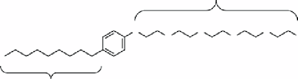
Hydrophilic region
O
O
O
O
O
OH
Hydrophobic region
fIGure 16.28
Structure of the surfactant Igepal CO-520.