Chemistry Reference
In-Depth Information
(a)
(b)
99m
Tc-DPA-ale-endorem
99m
Tc-DPA-ale
160
30
120
25
20
L
80
15
10
40
S
5
0
0
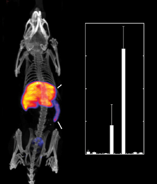
fIGure 16.26
Whole-body SPECT-CT maximum intensity projection (left) and biodistribution studies (right) of
99m
TcDPA-ale-
Endorem (a) and
99m
TcDPA-ale (b). Reprinted with permission from Ref. [87]. Copyright 2011 American Chemical Society. (
See insert for
colour representation of the figure.)
)
To form a tumour, Cli36-sshBirA cells were injected intracranially in mice. Streptavidin with AlexaFluor750 was used
for fluorescence-mediated tomographic imaging. Subsequently, streptavidin labelled with either
111
In-DTPA-biotin or
SPIONs were injected intravenously for SPECT imaging or MRI imaging respectively. The tumour was detected using
bioluminescence (following injection of coelenterizine), fluorescence tomography, SPECT, and MRI.
A novel targeted MRI-CT nanoparticle system was investigated using FePt alloy particles and the HER2 antibody [91].
These nanoparticles were synthesised by heating Pt(acac)
2
and Fe(CO)
5
with dioctyl ether, hexadecandiol, and oleyl amine
or oleic acid. The size was found to be 3, 6, or 12 nm upon varying the heating conditions [91]. The surface was passivated
by coating with cysteamine, and the amine groups were used for peptide coupling to the HER2 antibody for targeting. The
particles were cuboctahedral at 3 and 6 nm, and cubic at 12 nm. The ratios of Fe:Pt were 58:42, 51:49, and 33:67 respectively.
Cell viability with MTT showed ~90% up to 10 mM Fe. Extensive biodistribution studies were undertaken at several time
points. In general, the highest concentrations of the particles were found in the lungs and spleen. Importantly, the particles
were found in the brain, indicating that these small particles may be useful for brain imaging.
Imaging using CT and MRI showed that only the 12 nm particles were appropriate for MR imaging, because they were
comparable to Resovist at 3 T. 10 mM in Fe was needed to detect enhancement in the image. As a CT agent, the 12 nm par-
ticles showed similar contrast to the same concentration of commonly used iodine agents. In cell pellets, the MRI and CT
showed greater labelling of MBT2 (HER2+) cells than MBT2-KD cells.
In vivo
, tail vein injection of the 12 nm particles in
mice with transplanted MBT2 tumours allowed the visualisation of the tumour by both MRI and CT.
Nanochains of iron oxide particles were used for geometrically enhanced multivalent docking [92]. The nanochains were
formed by a previously reported multistep process [93]. First, using solid phase chemistry, amine-functionalised SPIONs
(N-SPIONs) were covalently bonded to a surface via DTSSP (Scheme 16.12). This attachment allows the SPIONS to be freed
by reduction of the S-S bond, leaving the particles with amine (NH
2
) groups on one side and thiol (SH) groups on the other side
(N,S-SPIONs). In the second step, N,S-SPIONs were again combined with DTSSP beads. The amine groups react with the
exposed SulfoNHS and leave the -SH groups exposed. These are reacted with Sulfo-SMCC, which contains a maleimide group
and a sulfoNHS group. The thiol groups react with the maleimide to form a highly stable thioether bond. N,S-SPIONs are
added and the amine groups react with the sulfoNHS group. This step is repeated n times to make a chain of n + 2 particles.