Chemistry Reference
In-Depth Information
The use of phosphonate groups has proven to be a stable method of nanoparticle surface modification [86]. In this work,
the metal-binding chelate dipicolyl amine (DPA) was combined with alendronate (ale) in order to introduce
99m
Tc chelates
onto the surface of SPIONS [87]. First,
99m
Tc -DPA-ale (Figure 16.24) was formed by mixing
99m
Tc(CO)
3
(H
2
O)
3
+
with
DPA-ale in water with brief heating [88]. Finally,
99m
TcDPA-ale was heated with Endorem, a commercially available
dextran-coated SPION. The transverse relaxivity was found to be r
2
~ 11 at 9.4 T. SPECT, CT, and PET were used to show
that the
99m
TcDPA-ale-Endorem particles accumulated in the liver and spleen, in contrast to free
99m
TcDPA-ale, which
localised in the femur (Figures 16.25 and 16.26).
16.6.2
targeting
Streptavidin (SA) is a tetrameric protein 66 kDa in size that binds to the small molecule biotin. Each of the four binding sites
is identical with the highest binding constant of known non-covalent interactions. Streptavidin serves as the 'nanoparticle'
for a NIRFI/SPECT/CTsystem [89]. First, Cy5.5-NHS was attached to biocytin and combined with SA in a 1:1 ratio. Biotin-
DOTA was added to the Cy5.5-SA in a 1:1 ratio and added to a solution of
111
InCl
3
. Finally, the HER2 antibody was biotinyl-
ated according to literature procedures and added to the SA. The agent was administered to mice intravenously. Fluorescence
at 2, 15, and 40 h as well as SPECT and CT showed accumulation in the liver, kidneys, spleen, and tumour. SUM190
(HER2+) xenograft tumours accumulated the agent, whereas SUM149 (HER2-) tumours did not.
A multimodal system based on the biotin-avidin interaction has been developed for the detection of brain tumours. The
Gaussia luciferase reporter gene was fused with a membrane anchor and BAP, biotin acceptor peptide. The biotin acceptor
peptide was attached so that biotin ligase attaches biotin on the cell surface at a specific lysine residue [90]. This approach
allows the cells to be detected with any modality attached to streptavidin (SA).
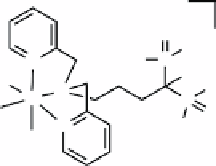
+
O
OH
OH
OH
O
HO
P
N
OC
N
Tc
P
OC
CO
N
HO
fIGure 16.24
Structure of bisphosphonate chelate
99m
TcDPA-ale.
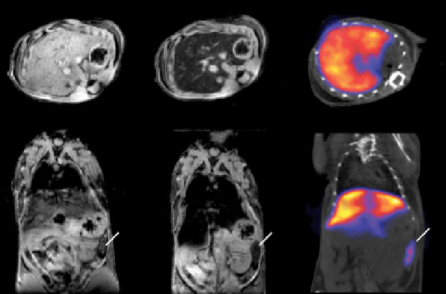
(a)
(b)
(c)
L
S
L
S
S
L
fIGure 16.25
Short-axis view (top) and coronal view (bottom) images: (a)
T
2
*-weighted MR images before injection of
99m
TcDPA-
ale-Endorem, (b)
T
2
*-weighted MR image 15 min. post injection, and (c) nanoSPECT-CT image of the same animal in a similar view
45 min. post injection. Contrast in the liver (L) and spleen (S) changes after injection due to accumulation of
99m
TcDPA-ale-Endorem, in
agreement with the nanoSPECT-CT image, which shows almost exclusively liver and spleen accumulation of radioactivity. Reproduced
with permission from Ref. [87]. (
See insert for colour representation of the figure.)
)