Chemistry Reference
In-Depth Information
structures, which was corroborated by a follow-up study [88]. although accurate comparison between the two
modalities was not feasible because fluorescence imaging was not very quantitative, such proof-of-principle study did
demonstrate for the first time the direct comparison of molecular optical and planar nuclear imaging for subcutaneous and
deeper tumours.
The c(RgDyK) peptide has also been labelled with
111
In and the NIRF dye cypate and investigated in a 4T1 murine breast
cancer model [89]. With tumours visualised by both optical and nuclear imaging methods in a receptor-specific manner, it
was suggested that such a dual-modality imaging approach could provide important complementary diagnostic information
for improving patient management.
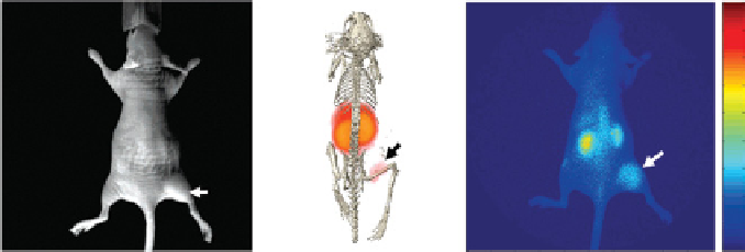
The cytokine interleukin (Il)-11 and its receptor, Il-11Rα, have been linked to breast cancer development and progres-
sion, as well as bone metastasis [90-92]. a cyclic peptide that mimics the receptor binding motif within Il-11, c(CgRRaggSC),
has been identified through phage display [93, 94]. a dual-modality imaging agent,
111
In-DTPa-Bz-NH-Sa-K(IR-783-S-Ph-
CO)-c(CgRRaggSC)NH
2
, was constructed in which DTPa and a fluorescence dye (IR-783-S-Ph-CO) were linked to the
peptide via a lysine linker [95]. The selection of IR-783-S-Ph-CO-NHS (NHS denotes N-hydroxysuccinimide) as the fluoro-
phore over its analogues was due to its superior chemical stability [96]. Cell-based studies revealed that the cyclic peptide
maintained the targeting capability to Il-11Rα after conjugation of both the optical and radioisotope labels. Cross-validation
and direct comparison of optical and nuclear imaging of the agent in a small animal tumour model was achieved using a
single injection, which demonstrated the tumour-targeting capability of the dual-modality probe
in vivo
(Figure 15.8).
(a)
SO
3
H
SO
3
H
+
N
S
Ala
Arg
Gly
Gly
Arg Gly
Ser
NH
H
O
N
O
HN
S-S
CONH
2
O
HN
O
N
O
N
N
In
O
O
O
O
O
O
O
O
(b)
White light
SPECT/CT
Optical
×10
4
2
1.8
1.6
1.4
1.2
1
0.8
0.6
0.4
0.2
Tumour
Tumour
Tumour
FIgure 15.8
Dual-modality SPECT/optical imaging of Il-11Rα. (a) Chemical structure of the dual-modality probe. (b) SPECT/CT
and optical images of a tumour-bearing mouse at 24 h after probe injection. adapted with permission from Ref. [95]. (
See insert for colour
representation of the figure.)
)