Chemistry Reference
In-Depth Information
Another novel multimodality imaging technique was recently demonstrated by Lin and coworkers. They fabricated a
Fe
3
o
4
@NagdF
4
:Yb,Er@NagdF
4
:Yb,Er core-multishell structure to combine UCL and T
1
/T
2
-weighted mRI together [99].
13.4.5.2 In Combination with X-ray CT Imaging
X-ray CT is another conventional imaging technique in the clinic. The
CT image is based on the different X-ray absorption abilities of tissues. The common CT contrast agents consist of heavy
elements, such as iodine and gold atoms. Lanthanide elements are located in the f-block (the sixth period) of the periodic
table, and the large atomic numbers of lanthanide elements endow them with large absorption coefficiency for the X-ray
techniques. Therefore, most lanthanide-based UCNPs can be used directly as CT contrast agents.
Cui and co-workers investigated the X-ray attenuation ability of Yb,Er/Ho/Tm co-doped NagdF
4
UCNPs [100]. After
subcutaneous injection into mice, UCNPs generated both UCL and CT signals. Lu and co-workers employed NaYbF
4
as the
host material for the UCL and CT imaging [101]. The PEg-coated NaYbF
4
:gd,Yb,Er nanoparticles showed good CT con-
trast effects comparable to the commercial CT contrast agents. These works provide the evidence that UCNPs can serve as
CT contrast agents and showed different metabolism characteristics compared to commercially used CT contrast agents.
13.4.5.3 In Combination with Positron Emission Tomography (PET) and Single-Photon Emission Computed Tomography
(SPECT) Imaging
Positron emission tomography (PET) and single-photon emission computed tomography (SPECT) techniques
can give accurate 3D information about the distribution of radioactive elements.
18
F is a commonly used radionuclide for PET
imaging. Due to the high affinity between rare earth and fluorine ions, fluoride ions can directly bind to the surface of UCNPs. Li
and co-workers proved that >90% of
18
F
−
was bound to the UCNPs after mixing them in an aqueous solution and shaking for 5 min
(Figure 13.11) [102]. This convenient method provides a powerful tool to investigate the biodistributions of UCNPs via PET imaging.
(a)
Injection site
Lymphatic circulation
Lymphatic vessel
Lymphocytes
18
F
-
18
F
-
labelling of RE materials
Sentinel lymph nodes
(b)
(c)
80
70
Lymph node
60
50
R
L
Na
18
F
40
18
F
-
NaYF
4
NPs
30
20
10
0 0 0 0
Time after injection (min)
40
50
60
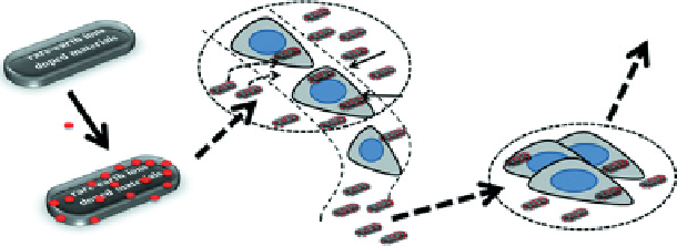
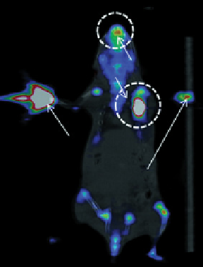
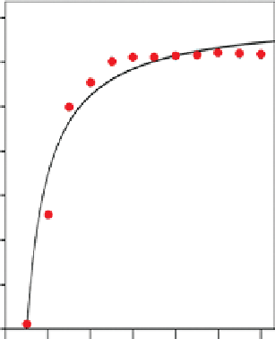
FIgure 13.11
(a) Schematic representation of the preparation of
18
F-labelled UCNPs and the lymph node imaging mechanism. (b)
PET/CT imaging of lymph node 30 min. after subcutaneous injection of 740 kBq/0.05 mL
18
F
-
UCNPs into the left paw footpad. Na
18
F
was injected into the right paw as a control experiment. (c) Curvilinear regression of real-time PET quantification detection of lymph node
signals. Reprinted with permission from Ref. [102]. Copyright 2011 Elsevier Ltd. (
See insert for colour representation of the figure.)
)