Chemistry Reference
In-Depth Information
(b)
T
1
-MRI
(d)
T
2
-MRI
(a)
(c)
(e)
[Gd]: 0.30 mM
[Er]: 0.86 mM
(f)
3.0 W
Zero
[Si]: 129.8 mM
Laser power (W)
(g)
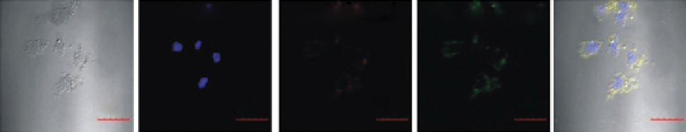
Bright eld
DAPI
Red channel
Green channel
Merged
50 um
50 um
50 um
50 um
50 um
(h)
(j)
(l)
Tumour
Tumour
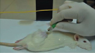
980 nm Laser
Tumour
Pre-injection
T
1
-MRI
T
2
-MRI
Pre-injection
Pre-injection
(i)
(k)
(m)
Tumour
Tumour
Tumour
T
1
-MRI
T
2
-MRI
Post-injection
Post-injection
Post-injection
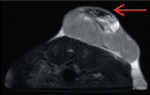
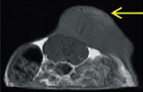
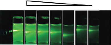
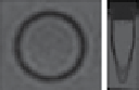
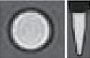
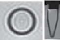
FIgure 13.10
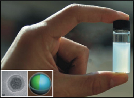
(a) Digital photograph of water soluble silica-protected NagdF
4
-based UCNPs. T
1
-mR images of (b) pure water (r
1
=
0.35 1/s) and (c) UCNPs (0.2 nm) (r
1
= 2.03 1/s). T
2
-mR images of (d) pure water (r
2
= 6.05 1/s) and (e) UCNPs (r
2
= 50 1/s). (f) Digital
photos of samples under varied NIR laser powers. (g) Confocal images of mCF-7 cells incubated with the probe.
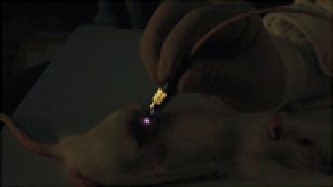
In vivo
optical imaging
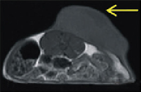
in tumour: (h) pre-injection, (i) post-injection.
In vivo
T
1
-mR images of tumour: (j) pre-injection, (k) post-injection.
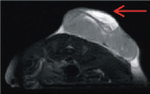
In vivo
T
2
-mR images
of tumour: (l) pre-injection, (m) post-injection. Tumour sites were marked with yellow and red arrows. Reprinted with permission from
Ref. [91]. Copyright 2011 Wiley-vCH verlag gmbH & Co. KgaA, Weinheim. (
See insert for colour representation of the figure.)
)
Recently, Li et al. have synthesised NaYF
4
:Yb,Tm@Fe
x
o
y
core-shell nanocrystals directly via an epitaxial growth method
[98]. A Fe
x
o
y
shell with 5 nm thickness provides T
2
-enhanced mRI characteristics. Lymphatic systems of small animals were
chosen as the model to demonstrate UCL/mRI dual-modality imaging. In addition, Liu and coworkers have developed an
electrostatic adsorption-based method to fabricate Fe
3
o
4
-UCNPs heterostructures. The UCL/mRI imaging was demon-
strated by tumour targeting and lymphatic imaging in small animals.