Chemistry Reference
In-Depth Information
R
1
R
1
R
2
R
2
N
N
N
N
R
3
R
3
N
N
CO
2
H
HO
2
C
500 μm
250 μm
125 μm
0 μm
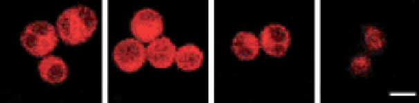
FigUre 12.26
Ligand utilised for the bimetallic helicate [Eu
2
L
3
] (R
1
= H, R
2
= Me, R
3
= PEg chain). Cells were incubated in the
presence of different concentrations of the helicate in RPMI-1640 for 24 h. The images were taken using a Zeiss LSM 500 META confocal
microscope (λ
ex
405 nm). (
See insert for colour representation of the figure.)
)
(a)
(b)
(c)
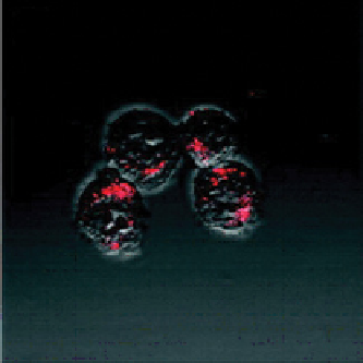
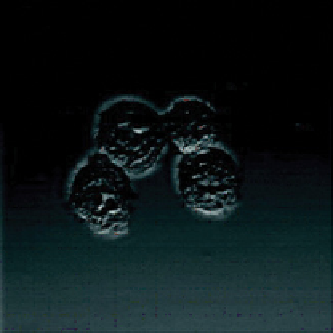
FigUre 12.27
Two-photon microscopy images of HeLa cells incubated with 200 μM of a bimetallic Eu
III
helicate [Eu
2
(L)
3
] in RPMI-1640
culture medium for 12 h at 37 °C, 5% CO
2
: (a) bright field image; (b) luminescence (λ
ex
= 750 nm, λ
em
= 570 − 650 nm); (c) merged image.
(
See insert for colour representation of the figure.)
)
probes, suggesting that these complexes are less advantageous in terms of assessing cell functionality through organelle
targeting. However, the intrinsic chirality of these bioprobes will also lend itself to potential application with CPL analyses.
It should also be noted that the complexes are very amenable to multi-photon excitation, wherein both two-photon
(Figure 12.27) and (uniquely) three-photon absorption have been demonstrated (i.e., excitation using NIR wavelengths) in
an imaging context [70].
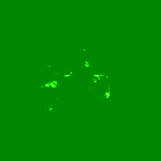
Two-photon imaging has also been shown with an architecturally simple Tb
III
complex, based upon a tripodal ligand
possessing a benzamide-type chromophore (Figure 12.33) with the formula [TbL(NO
3
)
3
]. Although the solid-state charac-
terisation of the complex revealed a polymeric structure, in solution the complex was sufficiently emissive (Φ
MeOH
= 0.11)
and possessed a long millisecond lifetime (in HEPES) indicative of Tb
III
-centred luminescence. Cellular studies were con-
ducted with A549, HeLa, and HONE1, each of which was exposed to the complex at 20 µg/ml concentrations for different
time durations; all showed good uptake of the complex. For the imaging studies, neither the free ligand nor the complex
absorbs light at 400-800 nm, but excitation at 800 nm induces Tb
III
-centred emission at 480 nm, indicative of a multi-photon
process, which was subsequently confirmed via power-dependence experiments. The cellular imaging (Figure 12.28)
showed that the green Tb
III
signal was predominantly localised in the cytoplasm, but not the nuclei. Assessments of the
cytotoxic effect of the complex (MTT assay) showed that at the concentration used for imaging the cell lines were viable
over a 24 hr period [71].