Biomedical Engineering Reference
In-Depth Information
models, a step necessary to determine whether
model predictions are borne out.
bone to adapt to its dynamic environment;
hence cells must be considered in any model of
engineered bone tissue. Even if “just” a scaffold
is modeled, it is necessary to remember that the
scaffold provides a surface for cell adhesion,
migration, and proliferation. The properties of
the scaffold will determine the maximum
number of cells residing in and on the scaffold
as well as the cells' mechanobiological milieu.
Third, although idealizations are made in
model development at one particular length
scale, the
implications of these idealizations at
other length scales
need to be addressed.
Fourth, any model must
incorporate more than
one function
, such as the interplay between the
mechanical role of bone and its structural
organization. This mandates a transdisci-
plinary approach to modeling bones in silico.
10.4 Computational Cell and
Tissue Models at Multiple
Length Scales
When bone is remodeled as a system, four
themes need to be addressed. First,
structure
and function of bone are interdependent
and
one cannot be addressed without affecting the
other; this is true across length and time scales
throughout the life of the animal. Second,
cells
are the living component of bone
, and it is the
movement and activity of cells that enables
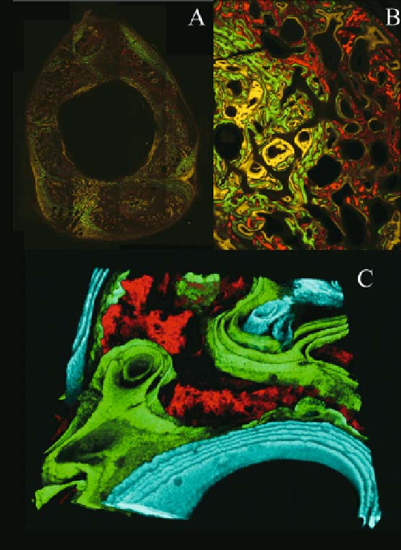
Figure 10.4.
Regeneration of bone
assessed by fluorochrome integration.
Fluorochromes are fluorescent agents
that are injected intravitally, i.e., into the
living animal. The agents are integrated
(biochemically through chelation) into
the mineralized matrix at the time that
new bone is being laid down. They allow
for elucidation of the timing of bone
apposition when fluorochromes with
different excitation and emission spectra
(imparting different colors in the micro-
graphs) are administered at different
time points. For this particular case, the
time points are captured during the
regeneration of bone within a segmental
bone defect in the femur of a sheep. (A)
Cross section showing robust regenera-
tion in the previously empty space of the
defect zone. (B) Alizarin red was admin-
istered first, followed by calcein green (2
weeks later) and tetracycline (yellow, 2
weeks thereafter). It is likely that the dis-
organized woven bone that was first laid
down during the rapid proliferation
stage of healing was remodeled and
replaced by more organized lamellar
bone during the remodeling phase
(green and yellow in B, green and blue in
C). (C) Confocal imaging allows for addi-
tion of the third dimension, which reveals
in more detail the volume and time
course of bone generation in a particular
volume of interest.