Biomedical Engineering Reference
In-Depth Information
Optical imaging
(reflectance)
Optical imaging
(feature extraction)
D
box
=1.42
L
MRI
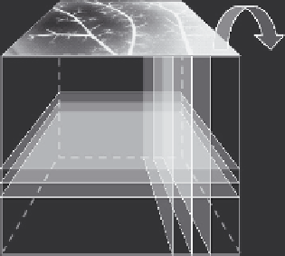
Fig. 2.9. Methodologicalpossibilitiesinexploring3Dspatialandtemporalcomplexityin
thebrain. The optical and MRI approaches can be instrumental in acquiring high resolu-
tion spatiotemporal data sets within the brain cortex (left panel) for biological functions,
such as supply networks of arteries and dynamic distribution of flow arriving through
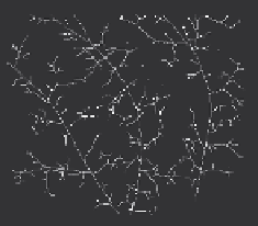
these networks to a 3D array of tissue microareas. Surface reflectance imaging with
feature extraction has proven adequate in assessing spatial complexity of the pial arte-
rial network in 2D in the rat (right panel). This network proved a spatial fractal that
distributes blood flow along the surface of the brain cortex via a self-similar pattern
of vascular arborization with a box dimension
(35)
, D
box
=1.42. This approach can
be taken further into the depth of the brain applied to high-definition 3D data sets of
structure and associated perfusion pattern in time. From a combination of the charac-
terization of complexity in spatial and temporal dynamics of tissue perfusion, a better
understanding of how global blood supply and local demand for adequate perfusion is
matched can emerge.
9. Summary
We have presented a overview of the fractal characterization of
spatial and temporal hemodynamic signals obtained from the
mammalian brain using non-invasive methods with reasonable
high temporal and/or spatial resolutions. We demonstrated that
the fractal analysis of optical (LDF, LSI, NIRS) and fMRI data can
capture the spatiotemporal complexity of cerebral hemodynamics.
Fractal analysis proved that the seemingly random fluctuations are
correlated according to the special order of self-similarity. We used
the Hurst exponent (
H
) and the spectral index (
β
) to characterize
the degree of correlation
(6, 14)
.
We showed in a rat model that the cortical perfusion is an
anti-persistent fractal process, which remains very stable during
hypotension
(16)
thus indicating strong self-organization
(23,24)
of regional flowmotion emerging from intrinsic segmental flow-
motion patterns. The capillary (parenchymal) and the small vessel