Biomedical Engineering Reference
In-Depth Information
A
12%
B
8%
4%
S1
BF
0%
P < 0.001
-4%
Trial 1 Rat 1
0
30
60
90
120
1.0
C
0.8
0.6
P < 0.01
0.4
0.2
0.0
-0.2
Trial 2 Rat 2
0
30
60
90
20
D
15
10
5
0
-5
Trial 3 Rat 3
0
30
60
90
Time (sec)
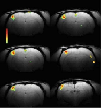
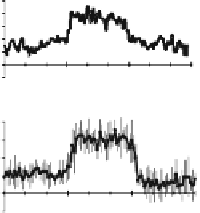
Fig. 10.5. Multi-modal responses from the contralateral whisker barrel field (S1
BF
)
during 8 Hz whisker stimulation in Sprague-Dawley rats. (
A
) Reproducibility of S1
BF
BOLD activation maps in the same subject (see Trial column; left) as well as other sub-
jects (see Rat column; right). The tmaps were generated by comparing the mean signals
in a 30-s baseline epoch before the stimulation. All data shown are from single trial runs.
Averaged time courses from the
S
1
BF
are shown for (
B
)BOLD(n= 6), (
C
) CBF (n= 5),
and (
D
)MUA(n = 5). The 30-s stimulation period is indicated by the black bar. Vertical
bars or gray shading represent standard deviations from the mean. (
See
Color Plate)
A
B
C
P < 0.001
S1
BF
P < 0.01
4 Hz
8 Hz
12 Hz
12%
12%
12%
8%
8%
8%
4%
4%
4%
0%
0%
0%
-4%
-4%
-4%
0
30
60
90
120
0
30
60
90
120
0
30
60
90
120
Time (sec)
Time (sec)
Time (sec)
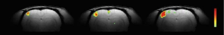
Fig. 10.6. Frequency-dependent BOLD responses from the contralateral whisker barrel field (S1
BF
) in Sprague-Dawley
rats. The t maps were generated by comparing the mean signals from 30-s epochs of baseline and stimulation. In the
same subject, S1
BF
BOLD responses are shown for (
A
)4Hz,(
B
) 8 Hz, and (
C
) 12 Hz whisker stimulations. The top and
bottom panels show the activation maps and time courses, respectively. All data shown are from single trial runs. The
30-s stimulation period is indicated by the black bar. (
See
Color Plate)
movement frequencies of less than 40 Hz (data not shown). Ros-
trocaudal movement of whiskers caused by air puffs over a wide
range of frequencies reproducibly stimulated the contralateral,
primary somatosensory area of the whisker barrel field (S1
BF
).
Repeated trials in the same subject and across subjects produced
activations in approximately the same locations in the contralat-
eral S1
BF
(
Fig. 10.5A)
. We observed medial to lateral extent of
contralateral S1
BF
activation, presumably because many whiskers