Biomedical Engineering Reference
In-Depth Information
demonstrate 3 Hz SWD activity, which is more comparable to
human SWD activity. Widespread increases were seen throughout
cortical and subcortical structures including the thalamus and hip-
pocampus. Unlike in GBL-induced seizures in rats, no significant
negative BOLD changes were seen although changes in the
hippocampus and the anterior cingulate were only seen during
shorter time points. As in the rat model, GBL-induced seizures in
monkeys most closely resembles status epilepticus
(90)
.
6.4. Discussion of
Animal Studies of
Spike-Wave
It is not yet clear why human fMRI studies of SWD show a mix
of cortical increases and prominent decreases while animal mod-
els with brief episodes of SWD show mainly cortical increases.
Similarly, it is unclear why prolonged SWD causes mainly BOLD
increases in the monkey GBL model, but both increases and
decreases in the rat GBL model. Some of this may reflect a lack of
understanding of the fundamental mechanisms of fMRI increases
and decreases during seizures. Direct recordings of neuronal
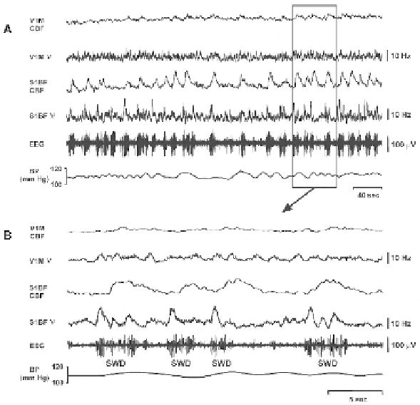
Fig. 9.5. CBF and neuronal activity recorded dynamically during SWD. (
A
) Combined
laser Doppler flowmetry and extracellular multiunit data recorded changes in CBF and
neuronal activity simultaneously during multiple episodes of SWD, along with EEG and
arterial blood pressure (BP) monitoring in a WAG/Rij rat under fentanyl-haloperidol anes-
thesia. (
B
) Parallel increases in CBF and neuronal firing rate (ν) in barrel cortex (S1BF),
and no or very small increases in primary visual cortex (V1M) induced by spontaneous
SWD. Panel (
B
) shows the data from the boxed region of panel A on an expanded time
scale. Reproduced with permission from Nersesyan et al 2004A, J Cerebral Blood Flow
Metab
(14)
.