Biology Reference
In-Depth Information
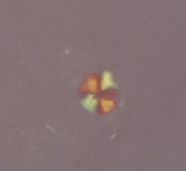
Fig. 1.
A CR-stained plaque viewed under polarized light. One gram of human brain was
homogenized and centrifuged at 4000 g for 2 min, and the pellet was washed and stained
with CR for two days. The amyloid plaque cores were further purified by sucrose gradient
centrifugation and filtration (40
m mesh filter). The plaque cores were dispensed on
a microscope slide and viewed under polarized light. The diameter of the plaque core is
around 15
µ
µ
m.
were at that time thought to be amorphous aggregates composed of starch,
but were later found to be ordered fibrillar deposits made of specific pro-
teins. At the beginning of the last century, the deposits were found to show
red-green bi-refringence in polarized light after staining with the dye
Congo red (CR) (Fig. 1). This phenomenon occurs when the CR molecules
are arranged in an ordered fashion, and bi-refringence is one criterion for
amyloid. Around 50 years ago, amyloid was subjected to electron
microscopy (EM) examination and found to be composed of thin fibrils
about 8 nm in diameter. When the amyloid fibrils were subjected to X-ray
diffraction, two sets of arcs could be observed, corresponding to distances
of 4.8 Å and 10-11 Å. These numbers reflect the distances between the
peptide strands and between the
β
-sheets, respectively, in a cross-
β
fiber.


Search WWH ::

Custom Search