Biomedical Engineering Reference
In-Depth Information
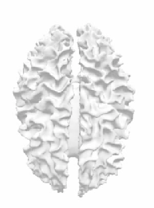
Figure 8.
Several steps in a surface evolution to cover the white matter of the brain from an
MRI dataset of size
. The top Figure shows initialization of the algorithmby
two spheres inside the white matter; the middle illustration depicts an intermediate step that
represents evolution; the bottom Figure represents the final result of the surface evolution.
See attached CD for color version.
256
×
256
×
124
cases; however, it will be still feasible to make a gross comparison of the volumes
of the dyslexic and normal brains.
The outer compartment is extracted by detecting the outer contour of the
white matter and moving it inward to obtain a boundary between the inner and
outer compartments. The reader may expect that moving inward for the same
distance in all cases may not result in a significant difference in volumes, so we