Biomedical Engineering Reference
In-Depth Information
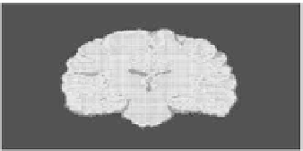
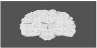
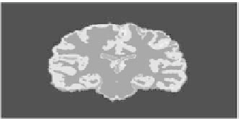
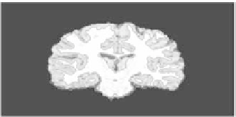
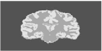
Figure 7.
Three steps in the evolution of the three classes of segmentation of a T1-weighted
MRI image in the coronal section.
Left column
: blue contour represents white matter
region, green contour represents gray matter, and the CSF is marked in red. Associated
adaptive regions are given in the right column. See attached CD for color version.
that we will be using here is the same as that discussed by Herbert et al. [18],
where the cerebral white matter was divided into inner and outer zones based on
a voxel-by-voxel basis according to an arbitrary distance from the white matter
boundary. The arbitrary choice of the distance in the present study does make
sense as long as it is sufficiently large to include the layer where communications
between the minicolumns take place. In other words, the way we define the outer
compartment of the white matter or the region of interest is the region where the
axon ends and the synapses communicate to different dendrites to transfer infor-
mation. Therefore, we have two choices to determine such a region: either to ask a
neuroscientist to draw this boundary (between the inner and outer compartments)
manually, which is very effort and time consuming, or to choose a distance that
will be sufficiently large so that it is guaranteed that the communications between
the minicolumns occur through it. Bearing in mind that if the chosen distance is
larger than the actual one “that could be determined by a neuroscientest,” this will
just add a bias to the volumes, but the differential comparison will still be valid.
In other words, the choice of distance will surely affect the volumes of individual