Biomedical Engineering Reference
In-Depth Information
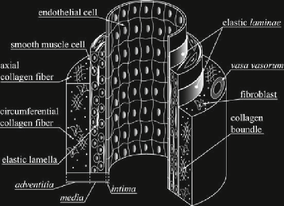
Fig. 7.1.
Structural anatomy of an artery. See [20] for more detailed pictures underlining peculiar
features of elastic and muscular arteries
gorized according to two general types: “elastic arteries”, including the aorta, main
pulmonary arteries, common carotids, and common iliacs, and “muscular arteries”,
which include the coronaries, cerebrals, femorals and renals. Elastic arteries tend to
be larger-diameter vessels located closer to the heart, whereas muscular arteries are
smaller-diameter vessels closer to the arterioles. Transitional arteries, such as the
external carotids, exhibit some characteristics of the elastic and muscular types [20].
Regardless of the type, all arteries consist of three layers: the
tunica intima
,
tu-
nica media
,and
tunica adventitia
(Fig. 7.1). The
intima
is similar in most elastic
and muscular arteries, typically consisting of a monolayer of endothelial cells and
an underlying thin (
80 nm) basal
lamina
. Exceptions include the aorta and coro-
nary arteries in which the
intima
may contain a subendothelial layer of connective
tissue and axially oriented smooth muscle cells. Endothelial cells are usually flat
and elongated in the direction of the blood flow; exceptions occur near bifurcations
wherein the blood flow is complex and the cells are often polygonal in shape. En-
dothelial cells are interconnected and may communicate via in-plane junctions or
with underlying smooth muscle cells via short processes that extend through the
basal
lamina
and into the
media
. The basal
lamina
consists largely of net-like type
IV collagen, the adhesion molecules laminin and fibronectin, and some proteogly-
cans; it provides some structural support to the arterial wall, but acts primarily as
an adherent meshwork on which the endothelial cells can grow. The
media
contains
smooth muscle cells that are embedded in an extracellular matrix of elastin and col-
lagen (primarily types I, III, and V) as well as an aqueous ground substance matrix
containing proteoglycans. Although the orientation and distribution of medial con-
stituents varies with species and location along the vascular tree, vascular smooth
muscle tends to be oriented helically, albeit nearly circumferentially in many vessels.
∼