Biomedical Engineering Reference
In-Depth Information
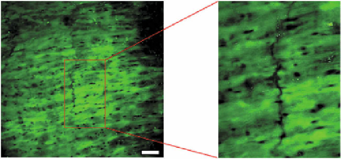
Fig. 6.10.
Multiphoton microscopy images from en face preparations of the basilar artery in a
human, fixed at 30 % strain, depicting autofluorescent elastin, by utilizing two-photon emission
(2PE) spectroscopy and revealing the fenestrated internal elastic lamina with a crack oriented in
the direction of applied load (circumferential direction, vertical in image). Bar = 50microns
years) individuals, and they are generally found in larger arteries, such as basilar and
internal carotid arteries, as transverse gaps 700-3000
m in length [47]. We are able
to assess these cracks using the UA-MPM system, Fig. 6.10.
Cracks have also been seen in experimental arteriovenous fistulas created be-
tween the common carotid artery and jugular vein [9]. In this latter case, no evidence
of elastolytic activity was found, so the cause was hypothesized to be due to direct
over-stressing (acute rupture) or from fatigue-type wear, discussed below. Histolog-
ical examination of the IEL from common carotid arteries subjected to longitudinal
[9] and circumferential [49] uniaxial failure tests have also shown damage to the
IEL in the form of mechanically-induced tears. Elastic fibres have been reported to
naturally fray and fragment over time [11]. As these fibres are progressively dam-
aged and possibly fail, the mechanical load will be transferred to the stiffer collagen
fibres [41, 92, 117, 119], leading to arterial stiffening.
μ
Sources of damage
Low cycle fatigue at high loads.
A study of the effect of balloon angioplasty on
bovine carotid arteries revealed that mechanically-induced damage under high loads
resulted in a shift in the mechanical response curve [92]. Elastic fibre damage was
not assessed histologically, but the results were applied to a continuum damage
model for arteries. Additionally, tests have been performed on ”dogbone” shaped
specimens from the human aorta to evaluate ultimate stress and extension ratio at
failure during circumferential and axial loading [79], though neither a continuum
damage framework nor histological techniques were employed. Scott et al. have
demonstrated damage in cerebral vessels under low cycle pressure inflation loading
conditions [102]. The tension/stretch curves displayed a loss of the toe region after
three cycles of loading to approximately 200 mmHg.