Biomedical Engineering Reference
In-Depth Information
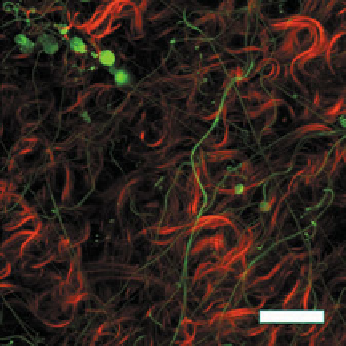
Fig. 6.9.
Adventitia of a rabbit carotid artery imaged with the UA-MPM system, revealing elastin
fibres (green) and collagen fibres (red), bar = 50
μ
m
= 566 kPa, and
γ
= 11.3. The thickness was 99
μ
m, width was 3.2 mm, initial length
of 3.7 mm.
This illustrative example was focused on the medial fibre distribution. A similar
approach is currently being used for a comprehensive study of the distinct contri-
butions of medial and adventitial collagen. Fig. 6.9 shows a representative image of
the adventitia obtained using the MPM-UA system.
6.4 Damage to the arterial wall
Mechanical damage can take the form of creation and growth of microvoids or mi-
crocracks. These are discontinuities in a medium that are often modelled as contin-
uous on a larger scale [69]. In the arterial wall, damage can occur to both the colla-
gen and elastin. However, while collagen naturally turns over in a health artery, the
mature arterial wall appears to be incapable of generating functional elastic fibres.
Further, damage to elastic fibres and the IEL are associated with a number of sig-
nificant diseases. For these reasons, much of the attention in this section is directed
at damage to the elastic fibres and the IEL. In this section, we discuss the physical
nature of elastin damage in the arterial wall as well as theoretical models of damage
to both collagen and elastic fibres/IEL. In Sect. 6.5, we consider the application of
these models to to cerebral angioplasty.
6.4.1 Physical nature of damage
In the case of the IEL, damage in the form of tears and fragmentation has been
reported. As early as the 1920s, Reuterwall reported tears in human IELs that have
been termed “Reuterwall's tears” by Hassler [47]. These are common in older (
>
30