Biology Reference
In-Depth Information
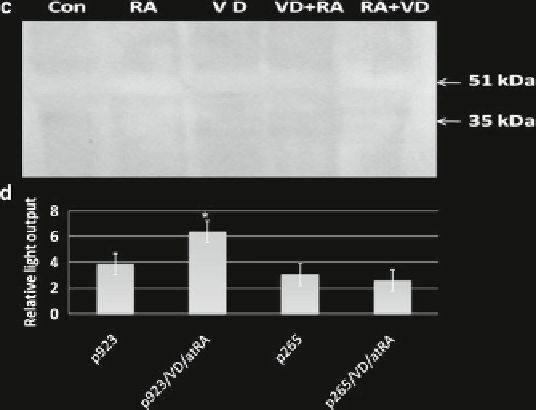
Fig. 27.2 (
continued
)
Deletion of regulatory region of the
TauT
promoter, which contains several tran-
scription factors (including two AP1 sites), abolished the effect of RA/1,25(OH)
2
D
3
on
TauT
expression. These findings suggest that the synergetic regulation of
TauT
expression by 1,25(OH)
2
D
3
and RA may require the formation of the RXR/VDR
complex, which in turn binds to the
TauT
promoter region to regulate
TauT
expres-
sion. Activation of RXR by RA appears to be critical for upregulation of
TauT
in
LLC-PK1 cells.
27.3.3
Regulation of TauT Expression by 1,25(OH)
2
D
3
and RA
in MCF-7 Cells
To study whether 1,25(OH)
2
D
3
and/or RA plays a role in
TauT
regulation in MCF-7
cells, the approaches used were similar to those described for the LLC-PK1 cells
experiments. As shown in Fig.
27.3
, 1,25(OH)
2
D
3
or atRA alone showed only a slight
effect on the expression of
TauT
. However, a combination of 1,25(OH)
2
D
3
and atRA
significantly decreased expression of
TauT
(as determined by reporter gene assay;
Fig.
27.3a
), taurine uptake (Fig.
27.3b
), and Western blot analysis (Fig.
27.3c, d
).
Interestingly, 9-cis RA showed little effect on
TauT
expression with or without
1,25(OH)
2
D
3
in MCF-7 cells (Fig.
27.3f
), suggesting that regulation of
TauT
by 9-
cis
RA or atRA occurs via different mechanisms in MCF-7 cells compared to LLC-PK1
cells. A summary of regulation of the
TauT
gene by 1,25(OH)
2
D
3
and RA in LLC-PK1
and MCF-7 cells is shown in Table
27.1
and further depicted in Fig.
27.4
.
Search WWH ::

Custom Search